Odontoid Fractures - Everything You Need To Know - Dr. Nabil Ebraheim
Dr. Ebraheim’s educational animated video describes fracture types of the odontoid process.
Most common fractures of the axis
Type I: Stable Avulsion Fracture of the alar ligament near the tip of the Odontoid. Treatment is soft collar, be aware of significant ligamentous injury.
Type II: Fracture at the base of the Odontoid Process. Most common and a troublesome fracture. Nonunion rate is 20-80% due to interruptions of the blood supply.
Type IIa: comminuted and unstable.
High nonunion rate in patients over 50 years of age and more than 5 mm of displacement.
Posterior displacement extension injury is rare (anterior displacement Is more common-flexion injury). Delay in treatment. Gap of more than 2 mm.
Treatment of type II fractures
•Halo jacket traction may be needed initially to reduce fracture.
•Keep halo for approximately 3 months
•30% nonunion rate in halo.
•Fusion C1-C2 (fusion decreases rotation by 50%). Odontoid screw is preferred in the young patient. Indications for fusion :
1-Nonunion
2-Displaced fracture in older patients
3- Failure of the halo
4-Type II a (comminuted/unstable).
Type III: Fracture through the body of C2. Rich blood supply so heals in the majority of the cases.
Treatment
•Cervical orthosis
•Halo if displaced
Diagnosis of odontoid fractures
•Lateral view
•Open mouth view
•CT scan is the best
Odontoid fractures in the elderly
•Simple fall, usually a missed diagnosis
•Usually associated with increased complications and mortality
•Do not use a halo, use external orthosis
•Fibrous union might be adequate if the fracture is not badly displaced, otherwise do fusion of C1-C2.
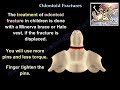
Odontoid fractures in pediatrics
Synchondrosis between odontoid and the C2 body, fusion by the age of six. Odontoid fractures occur in young children around 4 years of age.
Treatment
•Minerva brace
•Halo vest if displaced: use more pins and less torque. Finger tightness.
Differential diagnosis- Os Odontoidium: with oval shaped sclerotic edges and the os is smaller than the normal dens. In type II dens fracture: the odontoid shape is preserved with sharp edges. Os is smaller than normal odontoid and fixed to the C1 anterior ring. Os Odontoidium looks like a fracture, but it is probably an old trauma.
Become a friend on facebook:
http://www.facebook.com/drebraheim
Follow me on twitter:
https://twitter.com/#!/DrEbraheim_UTMC
Видео Odontoid Fractures - Everything You Need To Know - Dr. Nabil Ebraheim канала nabil ebraheim
Most common fractures of the axis
Type I: Stable Avulsion Fracture of the alar ligament near the tip of the Odontoid. Treatment is soft collar, be aware of significant ligamentous injury.
Type II: Fracture at the base of the Odontoid Process. Most common and a troublesome fracture. Nonunion rate is 20-80% due to interruptions of the blood supply.
Type IIa: comminuted and unstable.
High nonunion rate in patients over 50 years of age and more than 5 mm of displacement.
Posterior displacement extension injury is rare (anterior displacement Is more common-flexion injury). Delay in treatment. Gap of more than 2 mm.
Treatment of type II fractures
•Halo jacket traction may be needed initially to reduce fracture.
•Keep halo for approximately 3 months
•30% nonunion rate in halo.
•Fusion C1-C2 (fusion decreases rotation by 50%). Odontoid screw is preferred in the young patient. Indications for fusion :
1-Nonunion
2-Displaced fracture in older patients
3- Failure of the halo
4-Type II a (comminuted/unstable).
Type III: Fracture through the body of C2. Rich blood supply so heals in the majority of the cases.
Treatment
•Cervical orthosis
•Halo if displaced
Diagnosis of odontoid fractures
•Lateral view
•Open mouth view
•CT scan is the best
Odontoid fractures in the elderly
•Simple fall, usually a missed diagnosis
•Usually associated with increased complications and mortality
•Do not use a halo, use external orthosis
•Fibrous union might be adequate if the fracture is not badly displaced, otherwise do fusion of C1-C2.
Odontoid fractures in pediatrics
Synchondrosis between odontoid and the C2 body, fusion by the age of six. Odontoid fractures occur in young children around 4 years of age.
Treatment
•Minerva brace
•Halo vest if displaced: use more pins and less torque. Finger tightness.
Differential diagnosis- Os Odontoidium: with oval shaped sclerotic edges and the os is smaller than the normal dens. In type II dens fracture: the odontoid shape is preserved with sharp edges. Os is smaller than normal odontoid and fixed to the C1 anterior ring. Os Odontoidium looks like a fracture, but it is probably an old trauma.
Become a friend on facebook:
http://www.facebook.com/drebraheim
Follow me on twitter:
https://twitter.com/#!/DrEbraheim_UTMC
Видео Odontoid Fractures - Everything You Need To Know - Dr. Nabil Ebraheim канала nabil ebraheim
Показать
Комментарии отсутствуют
Информация о видео
Другие видео канала
 Odontoid Fractures - Everything You Need To Know - Dr. Nabil Ebraheim
 Jefferson Fracture - Everything You Need To Know - Dr. Nabil Ebraheim
 How to identify a vertebra (anatomy)
 Management of Odontoid Fractures
 The Patella (anatomy)
 Fracture Femur Types - Everything You Need To Know - Dr. Nabil Ebraheim
 Hangman's Fracture, C2 Fracture - Everything You Need To Know - Dr. Nabil Ebraheim
 Cervical Radiculopathy - Everything You Need To Know - Dr. Nabil Ebraheim
 Red Flags of Low Back Pain, When You Start to Worry -Everything You Need To Know -Dr. Nabil Ebraheim
 Supracondylar humerus fracture , pulseless hand - Everything You Need To Know - Dr. Nabil Ebraheim
 Vertebral ligaments
 Jones Fracture - Everything You Need To Know - Dr. Nabil Ebraheim
 Osteonecrosis Of The Hip Stages & Treatment - Everything You Need To Know - Dr. Nabil
 Fracture Of The Radial Head Essex Lopresti - Everything You Need To Know - Dr. Nabil Ebraheim
 Cervical Spine Injuries Jefferson Fracture - Everything You Need To Know - Dr. Nabil Ebraheim
 Galeazzi Fracture - Everything You Need To Know - Dr. Nabil Ebraheim
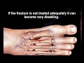 Stress Fractures Of The Metatarsal Bones - Everything You Need To Know - Dr. Nabil Ebraheim
 Ankle Fractures - Everything You Need To Know - Dr. Nabil Ebraheim
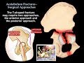 Acetabulum Fractures Surgical Approaches - Everything You Need To Know - Dr. Nabil Ebraheim
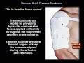 Humeral Shaft Fracture Treatment - Everything You Need To Know - Dr. Nabil Ebraheim