Jones Fracture - Everything You Need To Know - Dr. Nabil Ebraheim
Dr. Ebraheim’s educational animated video describes the condition of Jones fracture - 5th metatarsal base.
Follow me on twitter:
https://twitter.com/#!/DrEbraheim_UTMC
Jones Fracture
Sir Robert Jones (British Surgeon) sustained an acute fracture at the base of the fifth metatarsal bone while dancing, and the fracture was then named after him. The Jones fracture occurs at the metaphyseal/diaphyseal junction, and it extends into the intermetatarsal joint proximal to the metatarsocuboid joint. One joint articulates with the cuboid bone (metatarsocuboid) and the second joint (intermetatarsal) articulates with the 4th metatarsal. For the Jones fracture to be called “Jones Fracture”, the fracture must enter the intermetatarsal joint (fracture must be distal to the metatarsocuboid joint and must enter the intermetatarsal joint). The Jones fracture occurs about 1 ½ cm distal to the tuberosity of the 5th metatarsal bone. The 5th metatarsal bone is divided into the head and shaft. Jones fractures of the proximal fifth metatarsal occurs in the watershed area within 1.5cm of the tuberosity. The area where the Jones fracture occurs is an area of limited blood supply. There are multiple metaphyseal arteries in the tuberosity. Nutrient artery with intramedullary branches provides retrograde blood flow to the proximal fifth metatarsal. Fracture distal to the tuberosity will disrupt the nutrient artery supply resulting in relative avascularity. The Peroneus Tertius tendon is inserted into the dorsal metaphysis of the 5th metatarsal bone. The Peroneus Brevis tendon is inserted into the tuberosity of the 5th metatarsal bone. The plantar fascia is connected to the fifth metatarsal bone. When a Jones fracture occurs, the tendons will pull the fracture apart and prevent healing. This fracture could be mistaken for a sprain because a sprain is common on this side of the foot. There are three types of fractures at the proximal fifth metatarsal: Zone I, Zone II, and Zone III. Zone I fractures are avulsion fractures (pseudo Jones Fracture) and occur at the Peroneus Brevis insertion site; they require conservative treatment. Zone II fractures are acute fractures that occur at the metaphyseal-diaphyseal junction and involve the 4th and 5th metatarsal articulation. Zone III stress fractures are chronic fractures that occur distal to the 4th and 5th metatarsal articulation and may be associated with cavovarus foot deformity. In children, it is important not to make the wrong diagnosis of a fracture of the proximal 5th metatarsal base while looking at a normal growth plate. The growth plate is usually present between the ages of 9-14 years of age, and it is parallel and lateral to the metatarsal. X-rays will show the fracture and its location. An acute Jones fracture will have sharp margins with no intramedullary sclerosis. A stress fracture will have a wide fracture line with medullary sclerosis. With nondisplaced fractures, use a boot or a cast and be non-weight bearing for 6-8 weeks. 75% of fractures will heal. For athletes or a displaced fracture, do a screw fixation of the fracture (very popular technique). In the lateral view, the canal appears to be straight and narrow. In the AP view, the 5th metatarsal appears to be curved (lateral bow). Lateral bow of the 5th metatarsal may cause complications during surgery. There is vulnerability at the midshaft for perforation of the medial cortex. The canal is narrower in the dorsal plantar dimension, which is narrow in the lateral view. The point of entry for the wire or the screw is not centered. The fifth metatarsocuboid joint blocks the proximal canal projection, and this situation can cause complications. Each patients metatarsal should be evaluated individually for proper screw selection. Drill parallel with the shaft in the lateral plane and avoid the plantar direction. Avoid the sural nerve. You will probably need to use a 4.5 mm cancellous screw. The appropriate length of the screw that should be used is usually around 40-50mm. The diameter of the screw depends on the width of the canal. The screw threads must cross the fracture site. Failure of the procedure is attributed to poor blood supply or return of the athlete to activity before complete radiographic union.
Видео Jones Fracture - Everything You Need To Know - Dr. Nabil Ebraheim канала nabil ebraheim
Показать
Комментарии отсутствуют
Информация о видео
Другие видео канала
 Lisfranc Injury - Everything You Need To Know - Dr. Nabil Ebraheim
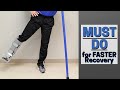 MUST Do Exercises with Injured Foot or Ankle- Faster Recovery
 Metatarsal Fracture Repair with Arthrex® Jones Screw
![5th Metatarsal Jones Fracture [Recovery, Treatment & Surgery] 2021!](https://i.ytimg.com/vi/8f0ECP1FtCc/default.jpg) 5th Metatarsal Jones Fracture [Recovery, Treatment & Surgery] 2021!
5th Metatarsal Jones Fracture [Recovery, Treatment & Surgery] 2021! Arthritis Of The Fingers - Everything You Need To Know - Dr. Nabil Ebraheim
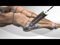 Metatarsal Fracture Repair with Arthrex® Hook Plate
 Neymar Jr Metatarsal Foot Injury | Doctor Explains Foot Fractures
 Spondylolysis, Spondylolisthesis, Spondylitis Spondylosis-Everything Need To Know-Dr. Nabil Ebraheim
 Nerve Injury Position of the Hand & Fingers - Everything You Need To Know - Dr. Nabil Ebraheim
 Dr. Frank Nisenfeld at MMI - Jones Foot Fracture
 Metatarsal Fractures | Complete Anatomy
 Physical Exam of the Lower Spine & Lower Extremity -Everything You Need To Know - Dr. Nabil Ebraheim
 Rotator Cuff Muscles - Everything You Need To Know - Dr. Nabil Ebraheim
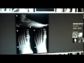 Fracture of the Fifth Metatarsal
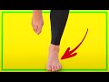 If You Have a Metatarsal Stress Fracture... WATCH THIS
 Diary of a Broken Foot | A Thousand Words
 Cauda Equina Syndrome - Everything You Need To Know - Dr. Nabil
 Stress Fracture Self Diagnosis for Runners by San Francisco Running Podiatrist.m4v
 Foot Fractures Fifth Metatarsal —Talking with Docs
 Fractures Of The Calcaneus - Everything You Need To Know - Dr. Nabil Ebraheim