Cervical Spine Injuries Jefferson Fracture - Everything You Need To Know - Dr. Nabil Ebraheim
Dr. Ebraheim’s educational animated video describes fractures types of the C1 cervical spine vertebrae, also called: Jefferson Fracture.
50% of patients will have associated spine injury. Canal is wide with low risk of spinal cord injury. These are difficult to visualize on x-rays. A “Junctional Fracture” could be missed. It’s a four part fracture with bilateral fracture of the anterior and posterior arch. There are variations which include two and three part fractures. Incomplete formation of the posterior arch can be mistaken as a fracture. At the upper cervical region, the spinal canal is 2.5 times larger than the cord size. At the upper cervical region, the spinal canal is 2.5 times larger than the cord size.
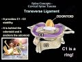
The atlantodental interval (ADI), should be less than 3mm in adults and less than 5mm in children. At approximately 3 to 5 mm A.D.I., there will be injury to the transverse ligament with an intact alar and apical ligament. An ADI greater than 5mm indicates injury to the transverse, alar, and apical ligaments. Bony injury with intact transverse ligament. Treatment consists of an orthosis for nondisplaced fractures and a halo jacket for displaced fractures. If an A.D.I is more than 3mm in adults, a fusion of C1-C2 may be necessary, even with bony avulsion of the transverse ligament. Lateral mass displacement more than 7mm. This is considered a significant injury with risk of spinal cord compression.
Type I Jefferson fractures have less than 7mm combined overhang with an intact transverse ligament. This is considered to be a stable fracture. Type II Jefferson fractures have more than 7mm combined overhang and a torn transverse ligament. This is an unstable. More than 7mm combined overhang.
A CT scan is the best imaging modality for diagnosing the bony injury. However, an MRI is the best in diagnosing a transverse ligament injury.
Become a friend on facebook:
http://www.facebook.com/drebraheim
Follow me on twitter:
https://twitter.com/#!/DrEbraheim_UTMC
Видео Cervical Spine Injuries Jefferson Fracture - Everything You Need To Know - Dr. Nabil Ebraheim канала nabil ebraheim
50% of patients will have associated spine injury. Canal is wide with low risk of spinal cord injury. These are difficult to visualize on x-rays. A “Junctional Fracture” could be missed. It’s a four part fracture with bilateral fracture of the anterior and posterior arch. There are variations which include two and three part fractures. Incomplete formation of the posterior arch can be mistaken as a fracture. At the upper cervical region, the spinal canal is 2.5 times larger than the cord size. At the upper cervical region, the spinal canal is 2.5 times larger than the cord size.
The atlantodental interval (ADI), should be less than 3mm in adults and less than 5mm in children. At approximately 3 to 5 mm A.D.I., there will be injury to the transverse ligament with an intact alar and apical ligament. An ADI greater than 5mm indicates injury to the transverse, alar, and apical ligaments. Bony injury with intact transverse ligament. Treatment consists of an orthosis for nondisplaced fractures and a halo jacket for displaced fractures. If an A.D.I is more than 3mm in adults, a fusion of C1-C2 may be necessary, even with bony avulsion of the transverse ligament. Lateral mass displacement more than 7mm. This is considered a significant injury with risk of spinal cord compression.
Type I Jefferson fractures have less than 7mm combined overhang with an intact transverse ligament. This is considered to be a stable fracture. Type II Jefferson fractures have more than 7mm combined overhang and a torn transverse ligament. This is an unstable. More than 7mm combined overhang.
A CT scan is the best imaging modality for diagnosing the bony injury. However, an MRI is the best in diagnosing a transverse ligament injury.
Become a friend on facebook:
http://www.facebook.com/drebraheim
Follow me on twitter:
https://twitter.com/#!/DrEbraheim_UTMC
Видео Cervical Spine Injuries Jefferson Fracture - Everything You Need To Know - Dr. Nabil Ebraheim канала nabil ebraheim
Показать
Комментарии отсутствуют
Информация о видео
Другие видео канала
 Cervical Spine Trauma - Everything You Need To Know - Dr. Nabil Ebraheim
 Jefferson fracture - radiology video tutorial (x-ray, CT)
 Ligaments of the Elbow Stability Of The Elbow - Everything You Need To Know - Dr. Nabil Ebraheim
 Jefferson Fracture - Everything You Need To Know - Dr. Nabil Ebraheim
 Cervical Myelopathy - What is it? How can we treat it?
 Unstable Subaxial C Spine Fractures: Anterior or Posterior - Daniel Gelb, MD
 Anatomy of a Cervical x-ray
 Pelvic Fractures - Everything You Need To Know - Dr. Nabil Ebraheim
 Piriformis Syndrome A Hidden Cause of Sciatica - Everything You Need To Know - Dr. Nabil Ebraheim
 Hangman's Fracture, C2 Fracture - Everything You Need To Know - Dr. Nabil Ebraheim
 Scaphoid Fractures - Everything You Need To Know - Dr. Nabil Ebraheim
 Proximal Humerus Fracture - Everything You Need To Know - Dr. Nabil Ebraheim
 Flexion Distraction Injury Of The lumbar Spine - Everything You Need To Know - Dr. Nabil Ebraheim
 Radiology of Spine Trauma
 Red Flags of Low Back Pain, When You Start to Worry -Everything You Need To Know -Dr. Nabil Ebraheim
 A Review Of Acetabular Fractures - Everything You Need To Know - Dr. Nabil Ebraheim
 Talus Fracture Types - Everything You Need To Know - Dr. Nabil Ebraheim
 Bone Tumors (Benign vs. Malignant)
 Odontoid Fractures - Everything You Need To Know - Dr. Nabil Ebraheim
 Spinal Cord injury , Examination & Evaluation - Everything You Need To Know - Dr. Nabil Ebraheim