Fracture Of The Radial Head Essex Lopresti - Everything You Need To Know - Dr. Nabil Ebraheim
Dr. Ebraheim’s educational animated video describes the condition radial head fracture - Essex Lopresti injury.
Follow me on twitter:
https://twitter.com/#!/DrEbraheim_UTMC
The Essex Lopresti injury affects the axial stability of the forearm. It is an injury to the interosseous membrane and the triangular fibrocartilage complex which could result in proximal migration of the radius. The Essex Lopresti fractures is difficult to diagnose, and the physician must restore the stability of the elbow and the DRUJ. The mechanism of injury is usually a fall onto an outstretched hand. The elbow will be in extension and pronation. There will be axial loading transmitted from the wrist to the radial head, which is combined with a valgus force, and this will create a fracture of the radial head. The radial head provides two types of stability: valgus stability and longitudinal stability. The radial head is secondary restraint to valgus load at the elbow, and it prevents proximal migration of the radius with some contribution from the interosseous membrane. Loss of this longitudinal stability occurs when the radial head fractures, plus injury to the DRUJ, and the interosseous membrane will become ruptured in this situation. In this situation, the radial head should be fixed or if the radial head is unreconstructable, replace the radial had by a radial head prosthesis, but never resect the radial head alone in this situation without replacing it. Radial head excision will result in proximal migration of the radius and ulnocarpal impingement with distal radioulnar joint instability. The problem is that not all hospitals are equipped with radial head prosthesis. You can excise the radial head if all ligaments are intact, but the problem is that you may not know that the DRUJ is involved. There are four types of radial head fractures. Type I is a nondisplaced fracture which has no block to forearm rotation. It has an early range of motion and does not require surgical treatment. Type II is a displaced fracture more than 2 mm. it requires fixation by screws or a plate. Type III is a comminuted fracture that is displaced or irreparable. It requires excision and prosthesis typically a metallic modular prosthesis. Excision alone can be done in some situations. Before you excise the radial head fracture, you must make sure all ligaments are intact, that you examined the patient and that there is no distal radioulnar joint (DRUJ) injury or elbow injury. If you have a patient with a comminuted radial head fracture, it is probably safer to replace it. Type IV fractures are associated with dislocation of the elbow joint. You should attempt to reduce the elbow joint with fixation of the fracture. This can be done with a plate or screws. Excision of the radial head and prosthetic replacement if the fracture is unreconstructable (cannot be repaired). Excision of the radial head alone is contraindicated in elbow dislocation or in Essex Lopresti fracture. To examine Essex Lopresti Fractures, you must first examine the DRUJ. Palpate the wrist for tenderness and excessive translation of the DRUJ. Examination of the DRUJ is very difficult; be sure to check the x-rays carefully. You may want to do the squeeze test, similar to what you do to check for high syndesmotic injury of the ankle and check if there is any tenderness there. You may want to get dynamic CT scans before surgery (it may show you some instability at the DRUJ. In surgery, you will do the radius pull test. More than 3mm of translation is concerning for longitudinal forearm instability (Lum & Trzeciak, 2018). Surgery for radial head fractures is done through posterolateral (Kocher) approach between the ECU and Anconeus muscles or through the lateral approach. Watch the safe zone for implant insertion to avoid impingement and loss of rotation. The radial head prosthesis usually I s cementless and acts as a stiff spacer until the ligaments heal, so it doesn’t have to be very snug into the canal (may fracture the proximal radius). You can use the modular system to check for the appropriate height. Make sure that you do not over stuff it. Visually assess widening of the lateral ulnohumeral joint, and also make sure that you are not blocking extension. You want to keep the lateral ulnar collateral ligament (ulnar humeral ligament) intact and stay above the equator of the radial head. When you stay above the equator in the radial head, it is less likely that you will injure the lateral ulnar collateral ligament (LUCL). Make sure that you understand the position of the posterior interosseous nerve which is about 4 cm. the posterior interosseous nerve crosses the proximal radius from anteriorly to posteriorly within the supinator muscle, 4 cm distal to the radial head. When you do the surgery, you want to pronate the forearm to protect the posterior interosseous nerve. Pronation pulls the nerve anteriorly away from the surgical field.
Видео Fracture Of The Radial Head Essex Lopresti - Everything You Need To Know - Dr. Nabil Ebraheim канала nabil ebraheim
Показать
Комментарии отсутствуют
Информация о видео
Другие видео канала
 Monteggia Fracture - Everything You Need To Know - Dr. Nabil Ebraheim
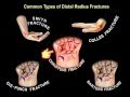 Common Types Of Distal Radius Fractures - Everything You Need To Know - Dr. Nabil Ebraheim
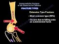 Supracondylar Fractures Of The Humerus In Children
 Elbow Anatomy Animated Tutorial
 Ligaments of the Elbow Stability Of The Elbow - Everything You Need To Know - Dr. Nabil Ebraheim
 What to expect after radial head fractures
 Lisfranc Injury - Everything You Need To Know - Dr. Nabil Ebraheim
 Arthritis Of The Fingers - Everything You Need To Know - Dr. Nabil Ebraheim
 Radial Head Fracture- Everything You Need To Know- Dr. Nabil Ebraheim
 Biceps Brachii Anatomy - Everything You Need To Know - Dr. Nabil Ebraheim
 Radius & Ulnar Shaft Open Reduction Internal Fix - Everything You Need To Know - Dr. Nabil Ebraheim
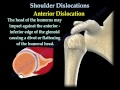 Shoulder Dislocations ,Everything You Need To Know - Dr. Nabil Ebraheim
 Cervical Spine Trauma - Everything You Need To Know - Dr. Nabil Ebraheim
 Coronoid Process Fracture - Everything You Need To Know - Dr. Nabil Ebraheim
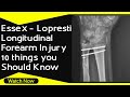 Essex - Lopresti Injury for FRCS
 Olecranon Fractures- Everything You Need To Know - Dr. Nabil Ebraheim
 Radius & Ulnar Shaft Fracture Approaches - Everything You Need To Know - Dr. Nabil Ebraheim
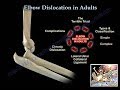 Elbow Dislocation In Adults - Everything You Need To Know - Dr. Nabil Ebraheim
 Essex-Lopresti fracture
 Humerus Fractures - Everything You Need To Know - Dr. Nabil Ebraheim