Supracondylar Fractures Of The Humerus In Children
Dr. Ebraheim’s educational animated video describing supracondylar fractures of the distal humerus in children.
Supracondylar fractures constitutes approximately 50% of all fractures.
The supracondylar region is thin and weak and thus it can fracture easily.
Fracture types:
1- Extension type:
- Most common type 95%
- Occurs due to falling onto an outstretched hand.
- The distal fragment is displaced posteriorly.
- Anterior interosseous neurapraxia is the most common nerve palsy occurring with supracondylar fractures.
- Injury to the anterior interosseous nerve will lead to weakness of the flexor digitorum prefundus muscle to the index finger, and the flexor pollicis longus muscle.
- The patient will not be able to do the OK sign or bend the tip of his index finger.
- Radial nerve neurapraxia is the second most common palsy and is evident by weakness of the wrist and fingers extension.
2- Flexion type:
- It is rare and occurs due to falling directly on a flexed elbow.
- The distal fragment is displaced anteriorly.
- This type of fracture may be accompanied with ulnar nerve neurapraxia.
- Injury to the ulnar nerve will lead to loss of sensation along the little finger.
- Later on, the patient may have weakness of the intrinsic hand muscles and clawing.
Gartland classification system:
- Type I: Nondisplaced
- Type II: Angulated with an intact posterior cortex
- Type III: Completely displaced
- Type IV: Complete periosteal disruption, and shows instability in flexion and extension.
Radiology:
- Plain AP and lateral x-rays should ne obtained.
- A posterior fat pad sign seen on a lateral view x-ray should increase your suspicion of an occult fracture around the elbow.
- The anterior humeral line:
• On a lateral view x-ray, the anterior humeral line is drawn along the anterior border of the distal humerus.
• Normally, the anterior humeral line should run through the middle third of the capitellum.
• In extension type fractures the capitellum will be displaced posteriorly relative to the anterior humeral ling.
- Baumann’s angel:
• Is formed by a line perpendicular to the axis of the humerus and a line going through the physis of the capitellum.
• normally, baumann’s angle should measure at least 11°.
Examination:
- on examination it is very important to assess the neurovascular structures.
- The anterior interosseous nerve is assessed by asking the patient to do the OK sign with his hand.
- The radial nerve is assessed by asking the patient to extend the wrist and the fingers.
- The ulnar nerve is initially assessed by loss of sensation along the little finger; later on the patient may have weakness of the intrinsic hand muscles and clawing.
Treatment:
- Non-operative treatment:
• Indicated for type I fracture.
• Usually consists of splinting or casting the elbow for duration of 3-4 weeks.
• It is very important to remember not to flex the elbow on the splint or cast more than 90° in order to avoid vascular compromise and compartment syndrome.
- Operative treatment:
• Type II and type III fractures are usually treated by closed reduction and percutaneous pinning.
• During reduction, pronation of the forearm during elbow flexion helps correcting a varus deformity.
- After reduction check for a gap in the fracture.
- The neurovascular bundle may be trapped there.
- Free the brachialis muscle from the fracture site if it is interapositioned there.
- Fixation is usually achieved with 2-3 divergent lateral pins, depending on stability.
- Medial pins can also be added depending on the stability.
- Open reduction is indicated only when closed techniques are unable to achieve appropriate reduction of the fracture.
- Avoid posterior dissection to preserve vascularity of the fractured segment.
- Fracture reduction and fixation should be done emergently in cases of vascular compromise.
Complications:
- Neurapraxia (ususally resolve and is thus observed).
- Cubitus varus deformity occurs due to malunion of the fracture.
- It only presents a cosmetic problem since it does not affect function.
- This deformity can be corrected later on by supracondylar valgus osteotomy.
- Vascular problems such as compartment syndrome.
- Volkmann’s ischemic contracture:
• Occurs due to compression of the brachial artery when the patient is placed in a cast in hyperflexion (more than 90°).
Important scenarios:
- Patient may present with a displaced type III fracture, and he has a pulseless hand.
1- He may have adequate circulation which is evident by a normal temperature and color of the hand.
2- Or he may have inadequate circulation, which is evident by a blue and cold hand.
- In both cases, urgent closed reduction and percutaneous pinning is required.
- After closed reduction and percutaneous pinning:
1- if the circulation is adequate: observe the patient and place in a splint that is 45°.
2- If the circulation is inadequate: the patient will require vascular exploration and repair.
Видео Supracondylar Fractures Of The Humerus In Children канала nabil ebraheim
Показать
Комментарии отсутствуют
Информация о видео
Другие видео канала
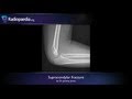 Supracondylar fracture - radiology video tutorial (x-ray)
 Supracondylar humerus fracture , pulseless hand - Everything You Need To Know - Dr. Nabil Ebraheim
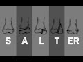 Salter Harris Fracture Classification - MEDZCOOL
 Colles Fracture Mnemonic
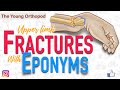 Named Fractures: UPPER LIMB, Fractures with EPONYMS, NEET PG, The Young Orthopod
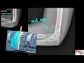 Elbow Xray Interpretation
 Brachial plexus
 Ligaments of the Elbow Stability Of The Elbow - Everything You Need To Know - Dr. Nabil Ebraheim
 Compartment syndrome - causes, symptoms, diagnosis, treatment, pathology
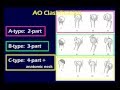 Proximal Humerus Fractures 1 (OTA lecture series III u03a)
 Common Types Of Distal Radius Fractures - Everything You Need To Know - Dr. Nabil Ebraheim
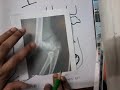 Supra condylar fracture of humerus
 Supracondylar Fracture Humerus & Circulation - Everything You Need To Know - Dr. Nabil Ebraheim
 Tibial Plateau Fractures - Everything You Need To Know - Dr. Nabil Ebraheim
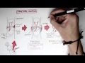 Bone Fracture - Types, Fracture Repair and Osteomyelitis
 Demonstration of supracondylar humerus fracture reduction in children... by Prof. G.K SINGH Sir
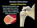 Shoulder Dislocations ,Everything You Need To Know - Dr. Nabil Ebraheim
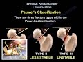 Femoral Neck Fracture Classification - Everything You Need To Know - Dr. Nabil Ebraheim
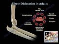 Elbow Dislocation In Adults - Everything You Need To Know - Dr. Nabil Ebraheim
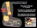 Supracondylar Fracture Of The Humerus In Children - Everything You Need To Know - Dr. Nabil Ebraheim