Anterior compartment of the leg - Muscles, vessels & nerves | Anatomy Tutorial
#legmuscles #anatomy #popliteal
Link for Donations https://paypal.me/studentlamedicina?locale.x=en_US
https://www.instagram.com/anatomy.knowledge/
The Tibialis anterior muscle lies against the lateral surface of the tibia.
Originates from the upper 2/3 of the lateral surface of the tibia and the adjacent interossous membrane.
Its insertion is on the medial cuneiform and the base of first metatarsal bone.
The extensor halucis longus muscle originates from the middle portion of fibula on the medial surface and the anjoining interosseous membrane. Its insertion is on the dorsal side of the base of the distal phalanx of the big toe.
The extensor digitorum longus muscle originates from the lateral condyle of tibia, anterior surface of fibula and adjoining interosseous membrane in its upper part. Its insertion is on the bases of middle and distal phalanges of the lateral four toes by four tendons.
The last muscle to be indicated is the peroneus tertius. This muscle originates on the distal anterior surface of the fibula and the adjacent interossous membrane. Its tendon inserts on the dorsal surface of the base of 5th metatarsal bone.
All the four muscles from the anterior compartment of the leg are supplied by the anterior tibial artery.
Видео Anterior compartment of the leg - Muscles, vessels & nerves | Anatomy Tutorial канала Anatomy Knowledge
Показать
Комментарии отсутствуют
Информация о видео
Другие видео канала
 Great Saphenous Vein & Small Saphenous Vein - Venous drainage of lower limb
 Sciatic Nerve - Anatomy Tutorial
 Femoral Artery and its branches - Anatomy tutorial
 Common peroneal nerve: Superficial peroneal nerve, Deep peroneal nerve : Anatomy Animations
 Muscles of Anterior compartment of Leg.
 Thigh Muscles - Medial Compartment of Thigh | Anatomy Tutorial
 Posterior compartment leg muscles
 Muscles of the Leg - Part 2 - Anterior and Lateral Compartments - Anatomy Tutorial
 Muscles of the Hand - Origin, Insertion, Nerve Supply | Anatomy Tutorial
 MUSCLES OF FRONT OF LEG | SIMPLIFIED ( Anterior Compartment #Clinical Anatomy)
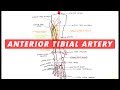 Anterior Tibial & Dorsalis Pedis arteries branches - Anatomy Tutorial
 Anatomy of Anterior Lateral Medial side of leg and dorsum of foot - Dr.G.Bhanu Prakash
 MUSCLES OF THE FRONT AND LATERAL SIDE OF LEG | ANATOMY | SIMPLIFIED
 Foot Anatomy Animated Tutorial
 Anatomy Of The Lower Leg - Everything You Need To Know - Dr. Nabil Ebraheim
 Tibial Nerve Anatomy Animation : Origin, Course, Branches, Tarsal tunnel syndrome
 Superficial Veins of Upper Limb - Basilic & Cephalic veins | Anatomy Tutorial
 Muscles of the Leg - Part 1 - Posterior Compartment - Anatomy Tutorial
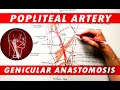 The Popliteal Artery branches & genicular anastomosis
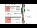 Anterior Leg Compartment | Origins, Insertions, & More