Tibial Nerve Anatomy Animation USMLE Step 1 : Origin, Course, Branches, Tarsal tunnel syndrome
►𝐉𝐨𝐢𝐧 𝐓𝐡𝐢𝐬 𝐂𝐡𝐚𝐧𝐧𝐞𝐥 𝐓𝐨 𝐆𝐞𝐭 𝐀𝐜𝐜𝐞𝐬𝐬 𝐓𝐨 𝐏𝐞𝐫𝐤𝐬 :- https://bit.ly/2RQHvTN
►𝐃𝐨𝐰𝐧𝐥𝐨𝐚𝐝 𝐭𝐡𝐞 𝐌𝐞𝐝𝐯𝐢𝐳𝐳 𝐚𝐩𝐩 𝐮𝐬𝐢𝐧𝐠 𝐭𝐡𝐞 𝐛𝐞𝐥𝐨𝐰 𝐥𝐢𝐧𝐤 👇👇👇👇 𝐃𝐨𝐰𝐧𝐥𝐨𝐚𝐝 👇👇👇👇
►𝐀𝐧𝐝𝐫𝐨𝐢𝐝 :- https://bit.ly/3ansFKq
📌𝐅𝐨𝐥𝐥𝐨𝐰 𝐨𝐧 𝐈𝐧𝐬𝐭𝐚𝐠𝐫𝐚𝐦 :-
https://www.instagram.com/drgbhanuprakash
Tibial Nerve Anatomy Animation: Origin, Course, Branches, Tarsal tunnel syndrome
-----------------------------------------------------------------------------------------------------
The tibial nerve is a major peripheral nerve of the lower limb. It has several cutaneous and motor functions in the leg and foot.
In this video, we shall look at the anatomy of the tibial nerve – its anatomical course, functions, and clinical correlations.
Nerve roots: L4-S3
------------------------------
Sensory: Innervates the skin of the posterolateral leg, lateral foot, and the sole of the foot.
Motor Innervates the posterior compartment of the leg and the majority of the intrinsic foot muscles.
Anatomical Course
-------------------------------
The tibial nerve is a branch of the sciatic nerve, and arises at the apex of the popliteal fossa. It travels through the popliteal fossa, giving off branches to muscles in the superficial posterior compartment of the leg. Here, the tibial nerve also gives rise to branches that contribute towards the sural nerve, which innervates the posterolateral aspect of the leg.
The tibial nerve continues its course down the leg, posterior to the tibia. During its descent, it supplies the deep muscles of the posterior leg.
At the foot, the nerve passes posteriorly and inferiorly to the medial malleolus, through a structure known as the tarsal tunnel. This tunnel is covered superiorly by the flexor retinaculum. Within this tunnel, branches arise from the tibial nerve to supply cutaneous innervation to the heel
Immediately distal to the tarsal tunnel, the tibial nerve terminates by dividing into sensory branches, which innervate the sole of the foot.
Motor Functions
---------------------------
The tibial nerve innervates the muscles of the posterior leg and the majority of the intrinsic foot muscles.
Posterior Compartment of the Leg
The muscles of the posterior compartment of the leg are organised into a superficial and deep compartment. They are all innervated by the tibial nerve.
Superficial:
Plantaris – plantarflexion of the ankle.
Soleus – plantarflexion of the ankle.
Gastrocnemius – flexion of the knee and plantarflexion of the ankle.
Deep:
Popliteus – “unlocks” the knee by laterally rotating the femur on the tibia.
Flexor hallucis longus – flexion of the great toe and plantarflexion of of the ankle
Flexor digitorum Longus – flexion of digits 2-5 and plantarflexion of the ankle.
Tibialis posterior – inversion of the foot and plantarflexion of the ankle.
Intrensic foot
The medial and lateral plantar branches of the tibial nerve provide innervation to all the intrinsic muscles of the foot (exept the extensor digitorum brevis, which is innervated by the deep fibular nerve).
Sensory Functions
------------------------------
In the popliteal fossa, the tibial nerve gives off cutaneous branches. These combine with branches from the common fibular nerve to form the sural nerve. This sensory nerve innervates the skin of the posterolateral side of the leg and the lateral side of the foot.
The tibial nerve also supplies all the sole of the foot via three branches:
Medial calcaneal branches: These arise within the tarsal tunnel, and innervate the skin over the heel.
Medial plantar nerve: Innervates the plantar surface of the medial three and a half digits, and the associated sole area.
Lateral plantar nerve: Innervates the plantar surface of the lateral one and a half digits, and the associated sole area.
Tarsal tunnel syndrome
Compression of the tibial nerve or its terminal branches will lead to an entrapment syndrome known as tarsal tunnel syndrome. This syndrome results in pain and sensory disturbances that affects the sole of the foot, and can cause palsies of the intrinsic foot muscles. This can also result from severe nerve trauma connected to fractures of the tibial shaft or medial malleolus.
#tibialnerve #tibialnerveanatomy #tibialnervecourse #tibialnervebranches #tibialnerveclinicalanatomy #tarsaltunnelsyndrome #tibialnervevideo #tibialnervelecture #tibialnerveanimation #tibialnerveinnervation
Видео Tibial Nerve Anatomy Animation USMLE Step 1 : Origin, Course, Branches, Tarsal tunnel syndrome канала Dr.G Bhanu Prakash Animated Medical Videos
Показать
Комментарии отсутствуют
Информация о видео
18 апреля 2021 г. 17:30:16
00:05:11
Другие видео канала
 Common peroneal nerve: Superficial peroneal nerve, Deep peroneal nerve : Anatomy Animations
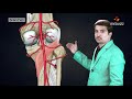 Posterior tibial artery : Origin , Course , Branches , Termination
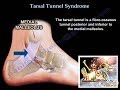 Tarsal Tunnel Syndrome - Everything You Need To Know - Dr. Nabil Ebraheim
 Obturator nerve Anatomy Animation : Origin, Course , Innervation and Clinical application
 Lower Extremity Nerve Injuries
 Popliteal Fossa
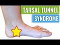 Tarsal Tunnel Syndrome Treatment & Diagnosis
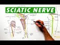 Sciatic Nerve - Anatomy Tutorial
 Tibial Nerve Entrapment - MSR Tibial Nerve Release Protocol
 The Sciatic Nerve Anatomy - Origin, Course, Relations, Branches, Distribution and Clinical anatomy
 Great saphenous vein - Animated Gross anatomy of lower limb ( Courtery : Dr vishram singh )
 ARCHES OF FOOT | ANATOMY | SIMPLIFIED
 The Popliteal Artery branches & genicular anastomosis
 Radial nerve Anatomy USMLE Origin, Course, innervation, Saturday night palsy, Wartenberg’s syndrome
 The Lateral Plantar Nerve - Everything You Need To Know - Dr. Nabil Ebraheim
 Femur Anatomy (Osteology) - General features , Attachments , Development #anatomy #MBBS #usmle #NMC
 Profunda femoris Artery or Deep femoral artery or The Deep artery of the thigh - Animation
 Ulnar nerve Anatomy: Origin, Course, Branches, Cubital Tunnel Syndrome || #Usmle videos
 Foot Drop, Peroneal Nerve Injury - Everything You Need To Know - Dr. Nabil Ebraheim
 Muscles in the Posterior Compartment of the Thigh / Hamstring Muscles