Common peroneal nerve: Superficial peroneal nerve, Deep peroneal nerve : Anatomy Animations
►𝐉𝐨𝐢𝐧 𝐓𝐡𝐢𝐬 𝐂𝐡𝐚𝐧𝐧𝐞𝐥 𝐓𝐨 𝐆𝐞𝐭 𝐀𝐜𝐜𝐞𝐬𝐬 𝐓𝐨 𝐏𝐞𝐫𝐤𝐬 :- https://bit.ly/2RQHvTN
►𝐃𝐨𝐰𝐧𝐥𝐨𝐚𝐝 𝐭𝐡𝐞 𝐌𝐞𝐝𝐯𝐢𝐳𝐳 𝐚𝐩𝐩 𝐮𝐬𝐢𝐧𝐠 𝐭𝐡𝐞 𝐛𝐞𝐥𝐨𝐰 𝐥𝐢𝐧𝐤 👇👇👇👇 𝐃𝐨𝐰𝐧𝐥𝐨𝐚𝐝 👇👇👇👇
►𝐀𝐧𝐝𝐫𝐨𝐢𝐝 :- https://bit.ly/3ansFKq
📌𝐅𝐨𝐥𝐥𝐨𝐰 𝐨𝐧 𝐈𝐧𝐬𝐭𝐚𝐠𝐫𝐚𝐦 :-
https://www.instagram.com/drgbhanuprakash
Common peroneal nerve ( Content from the textbook of anatomy by Dr vishram Singh )
----------------------------------------
The common peroneal nerve, also known as common fibular nerve, forms the lateral part of the sciatic nerve and supplies the leg.
Summary
origin: one of two terminal branches of the sciatic nerve
course: diverges laterally to enter the lateral compartment of the leg
terminal branches
-----------------------------
deep peroneal nerve
superficial peroneal nerve
motor supply: short head biceps femoris
sensory supply: cutaneous innervation of the posterolateral leg
Gross anatomy
-------------------------
Origin
One of two terminal branches of the sciatic nerve, with the division, typically occurring in the lower third of the thigh. It arises from the posterior division of the sacral plexus.
Course
Diverges away from the sciatic nerve by sloping medial and downwards to sit along the biceps femoris tendon where it passes through to the lateral aspect of the popliteal fossa. It exits the popliteal fossa over the plantaris and lateral head of gastrocnemius. Here the common peroneal nerve remains subfascial as it winds its way around the fibula neck to enter the lateral compartment of the leg where it can be felt and rolled in the living. It divides into its terminal branches - superficial and deep peroneal nerves.
Branches and supply
----------------------------------
muscular twigs to the short head of biceps femoris
cutaneous branches: fibular communicating branch, lateral sural cutaneous branch to innervate the posterolateral leg
terminal branches
#superficialperonealnerve #deepperonealnerve #commonperonealnerve #peronealnerve #lowerlimbnerves #anatomyanimations #animatedanatomy #nervesoflowerlimb #usmle #mbbs #medicalstudents #anatomyvideos #anatomylectures #anatomydrbhanuprakash #medvizz #proceum #usmleanatomyvideos #anatomylectures #peronealnerve #peronealnervevideo #peronealnerveexercises #peronealnervelecture #peronealnerveinjury
Видео Common peroneal nerve: Superficial peroneal nerve, Deep peroneal nerve : Anatomy Animations канала Dr.G Bhanu Prakash Animated Medical Videos
Показать
Комментарии отсутствуют
Информация о видео
10 октября 2017 г. 23:05:01
00:09:56
Другие видео канала
 Tibial Nerve Anatomy Animation USMLE Step 1 : Origin, Course, Branches, Tarsal tunnel syndrome
 Peroneal Nerve Entrapment - MSR Peroneal Nerve Release
 Great saphenous vein - Animated Gross anatomy of lower limb ( Courtery : Dr vishram singh )
 HIP Joint Anatomy Animation : Ligaments, Movements, Blood supply, Nerve supply and Hip Dislocation
 Common Peroneal Nerve - Deep Peroneal Nerve & Superficial Peroneal Nerve | Anatomy Tutorial
 Dorsalis pedis artery Animated lecture - ( Lower limb Gross anatomy from Dr vishram singh )
 Obturator nerve Anatomy Animation : Origin, Course , Innervation and Clinical application
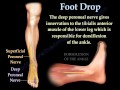 Foot Drop, Peroneal Nerve Injury - Everything You Need To Know - Dr. Nabil Ebraheim
 The Sciatic Nerve Anatomy - Origin, Course, Relations, Branches, Distribution and Clinical anatomy
 Common peroneal nerve- Chart | TCML
 Anatomy of Anterior Lateral Medial side of leg and dorsum of foot - Dr.G.Bhanu Prakash
 MUSCLES OF THE FRONT AND LATERAL SIDE OF LEG | ANATOMY | SIMPLIFIED
 ANATOMY OF FEMORAL TRIANGLE , FEMORAL CANAL , FEMORAL SHEATH
 Lower Extremity Nerve Injuries
 Radial nerve Anatomy USMLE Origin, Course, innervation, Saturday night palsy, Wartenberg’s syndrome
 Popliteal Artery - Anatomy, Branches & Relations
 Profunda femoris Artery or Deep femoral artery or The Deep artery of the thigh - Animation
 Femoral Nerve Anatomy: Origin, Course, Branches and Clinical application
 Anatomy of Hip Bone / innominate bone / Pelvis ( Osteology ): Ilium, Ischium, Pubis: Animation
 The Lumbar Plexus - Gross Anatomy