Femoral Nerve Anatomy: Origin, Course, Branches and Clinical application
FOLLOW ON INSTAGRAM:- https://www.instagram.com/drgbhanuprakash/
Channel Memberships: https://www.youtube.com/channel/UCG5TBPANNSiKf1Dp-R5Dibg/join
Femoral Nerve Anatomy Lecture: Gross Anatomy by Dr. G Bhanu Prakash
The femoral nerve is formed by the ventral rami of L2-L4, specifically the posterior divisions of the lumbar plexus.
The femoral nerve travels posterior to the inguinal ligament within the muscular lacuna. The muscular lacuna also contains the iliopsoas muscle.
NAVEL is a mnemonic for remembering the neurovascular structures that travel deep to the inguinal ligament into the femoral triangle.
N = femoral nerve
A = femoral artery
V = femoral vein
EL = empty space (femoral canal) and lymphatics
The femoral nerve provides motor innervation to the muscles of the anterior compartment of the thigh, with a few exceptions:
-the psoas portion of iliopsoas is innervated by muscular branches of the lumbar plexus
-tensor fasciae latae is innervated by the superior gluteal nerve
-pectineus is occasionally innervated by the obturator nerve
The femoral nerve provides sensory innervation to the skin of the anterior thigh and the anteromedial aspect of the leg. The afferent innervation of the leg is provided by the saphenous nerve (a branch of the femoral nerve that travels in the adductor canal).
Injury to the femoral nerve typically produces: weakness of hip flexion, loss of knee extension (no patellar reflex), and sensory loss over the territories described above. usmle videos
#femoralnerveanatomy #femoralnerve #femoralnervecourse #femoralnervebranches #femoralnerveanimation #femoralnervevideo #femoralnervelecture #femoralnerveanimated #femoralnerveexplained #femoralnerve #anatomyoffemoralnerve #mbbs #usmle #usmlestep1
Видео Femoral Nerve Anatomy: Origin, Course, Branches and Clinical application канала Dr.G Bhanu Prakash Animated Medical Videos
Показать
Комментарии отсутствуют
Информация о видео
29 июня 2017 г. 17:37:57
00:05:38
Другие видео канала
 Obturator nerve Anatomy Animation : Origin, Course , Innervation and Clinical application
 Great saphenous vein - Animated Gross anatomy of lower limb ( Courtery : Dr vishram singh )
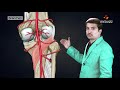 Posterior tibial artery : Origin , Course , Branches , Termination
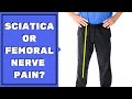 Is Leg Pain Sciatica or Femoral Nerve Pain? Must See to Assess & Stop Pain
 Femoral Nerve Anatomy - MBBS ANATOMY VIDEO LECTURES
 Sacral Plexus | Anatomy Tutorial
 The Lumbar Plexus - Gross Anatomy
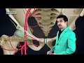 Femoral artery Anatomy : Origin , Course , Branches and Termination - Animated Lecture
 Femoral Nerve - Nerve Flossing - Kinetic Health
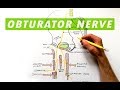 Obturator Nerve - Anatomy Tutorial
 Muscles of the Hip and Thigh - Human Anatomy | Kenhub
 Common peroneal nerve: Superficial peroneal nerve, Deep peroneal nerve : Anatomy Animations
 Dorsalis pedis artery Animated lecture - ( Lower limb Gross anatomy from Dr vishram singh )
 Ulnar nerve Anatomy: Origin, Course, Branches, Cubital Tunnel Syndrome, Guyon's canal syndrome
 ANATOMY OF FEMORAL TRIANGLE , FEMORAL CANAL , FEMORAL SHEATH
 Radial nerve Anatomy - Origin, Course, innervation, Saturday night palsy, Wartenberg’s syndrome
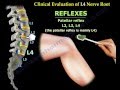 Femoral Nerve Anatomy - Everything You Need To Know - Dr. Nabil Ebraheim
 AXILLARY ARTERY ANATOMY ANIMATED LECTURE
 Sciatica Clinical Diagnosis - Everything You Need To Know - Dr. Nabil Ebraheim
 Femur Anatomy - General features , Attachments , Development , Fractures - MBBS , FMGE and NEET PG