Femoral Nerve Anatomy - Everything You Need To Know - Dr. Nabil Ebraheim
Dr. Ebraheim’s educational animated video describes the anatomy of the femoral nerve in a very easy and simple animation.
The femoral nerve arise from L2, L3, and L4 posterior divisions of the ventral rami. The anterior division of the ventral rami gives the obturator nerve. The femoral nerve is the largest branch of the lumbar plexus. The two muscular branches of the femoral nerve supply the psoas and iliac muscle (iliopsoas). the femoral nerve descends from the lumbar plexus in the abdomen through the psoas major muscle. The nerve then passes deep to the inguinal ligament where it then enters the femoral triangle of the thigh. Inside the femoral triangle, the nerve is lateral to the femoral artery. Distal to the inguinal ligament and under the Sartorius' muscle, the femoral nerve splits into the terminal branches. The branches are motor and sensory.
•Motor branches to Sartorius, Quadriceps and Pectineus.
•Sensory branches: intermediate femoral cutaneous nerve, saphenous nerve, medial femoral cutaneous nerve.
Motor function of the femoral nerve
Hip flexors
•Iliacus
•Psoas
•Pectineus
•Sartorius
Knee extensors
•Quadriceps femoris (includes four muscle): vastus lateralis, vastus intermedius, vastus medialis, rectus femoris (superficial layer).
The function of the femoral nerve is to provide motor innervation for the muscles that allow for flexion of the hip and extension of the knee.
Testing the motor functions of the femoral nerve: the function of the quadriceps muscle can be assessed with the patient being seated, the examiner applies resistance against leg extension. The function of the iliopsoas muscle can be tested by having the patient flex the hip against resistance.
Femoral nerve involvement, injury or palsy
Usually caused by trauma.
The manifestation of femoral nerve injury:
•Wasting of the quadriceps femoris
•Loss of knee extension
•Some loss of hip flexion (iliacus & psoas)
Differential diagnosis:
•Quadriceps tendon rupture
Other areas of interest for involvement of the femoral nerve
•Lumbar spine (lumbar disc herniation)
•Pelvis (pelvic hematoma)
Clinical evaluation of L4 nerve root
•Sensory area of L4
•L4: dorsiflexion of the ankle (tibialis anterior)
•Patellar reflex ( L2, L3, L4): the patellar reflex is mainly L4
•Femoral stretch test: positive test means the L3, L4 nerve roots are involved. Most disc herniations affect the L5-S1 nerve roots (use straight leg raise test). The test will stretch the femoral nerve and is positive when pain is felt on the ipsilateral anterior thigh.
The femoral nerve (saphenous nerve) may be affected with hematoma of the pelvis.
Muscles within iliopsoas compartment:
•Iliacus
•Psoas major
•Psoas minor
Hemorrhage in the pelvis and iliopsoas hematoma may usually be caused by
•Severe trauma
•Anti coagulation therapy
•Hemophilia or other blood diseases.
Clinical presentation: paresthesia around the medial side of the knee in the distribution of the saphenous nerve.
Become a friend on facebook:
http://www.facebook.com/drebraheim
Follow me on twitter:
https://twitter.com/#!/DrEbraheim_UTMC
Видео Femoral Nerve Anatomy - Everything You Need To Know - Dr. Nabil Ebraheim канала nabil ebraheim
Показать
Комментарии отсутствуют
Информация о видео
Другие видео канала
 Tests For Examination of the Lower Back - Everything You Need To Know - Dr. Nabil Ebraheim
 Femoral Nerve Anatomy: Origin, Course, Branches and Clinical application
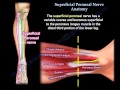 Superficial Peroneal Nerve Anatomy - Everything You Need To Know - Dr. Nabil Ebraheim
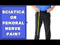 Is Leg Pain Sciatica or Femoral Nerve Pain? Must See to Assess & Stop Pain
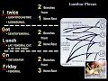 Lumbar Plexus - Everything You Need To Know - Dr. Nabil Ebraheim
 Sciatic Nerve Origin Variation & Course 1
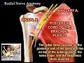 Radial Nerve Anatomy - Everything You Need To Know - Dr. Nabil Ebraheim
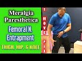 Meralgia Paresthetica! Femoral N Entrapment! FIX Thigh, Hip, & Knee Pain! ONE MOVE! | Dr Wil & Dr K
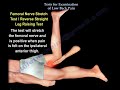
 Femoral Nerve Neurodynamic Tests
 Sciatic nerve: branches, course and clinical significance - Human Anatomy | Kenhub
 Low Back Pain - Disc Herniation ,Sciatica - Everything You Need To Know - Dr. Nabil Ebraheim
 Femoral artery Anatomy : Origin , Course , Branches and Termination - Animated Lecture
 Obturator Nerve Anatomy - Everything You Need To Know - Dr. Nabil Ebraheim
 Lumbosacral Plexus - Everything You Need To Know - Dr. Nabil Ebraheim
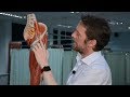 Nerves of the lower limb
 Femoral Nerve Anatomy - MBBS ANATOMY VIDEO LECTURES
 Rotator Cuff Tear ,injury - Everything You Need To Know - Dr. Nabil Ebraheim
 Sciatic Nerve Anatomy 3D - Everything You Need To Know - Dr. Nabil
 Femoral Nerve - Nerve Flossing - Kinetic Health
 Locating and Treating the Femoral Nerve - Neural Surface Anatomy Series - Stimpod NMS460