Obturator Nerve Anatomy - Everything You Need To Know - Dr. Nabil Ebraheim
Dr. Ebraheim’s educational animated video describes the anatomy of the Obturator nerve.
The obturator nerve is the second major nerve that arises from L2, L3 and L4 ventral rami of the lumbar plexus. The obturator nerve travels under the psoas major muscle and lies on the medial side of the psoas major. The obturator nerve then runs between the psoas and iliacus muscles through the pelvis towards the obturator foramen. The obturator foramen is covered by the obturator membrane.
The obturator externus muscle arises from the obturator membrane. Structures that run under the inguinal ligament:
The obturator canal is an opening in the obturator membrane at its superior lateral aspect. The obturator nerve is accompanied with vessels that exit the pelvis through the obturator canal. The obturator nerve and artery are at risk of injury from the placement of a retractor beneath the transverse acetabular ligament.
With an “open book” reduction, the reduction clamp is usually inserted through the medial side of the foramen away from the neurovascular bundle. Outside the foramen in the proximal part of the thigh, the obturator nerve divides into anterior and posterior divisions. The anterior division passes in front of the abductor brevis muscle and the posterior division passes behind the muscle.
Branches
Anterior division
•Adductor brevis
•Adductor longus
•Sensory branch
•Gracilis
Posterior division
•Obturator externus
•Adductor Magnus
One-third of the population will have an accessory obturator nerve arising from L3-L4. The accessory obturator nerve passes over the superior pubic rami and then dives below the pectineus muscle and joins the anterior division of the obturator nerve. The accessory obturator nerve supplies the pectineus muscle which is usually innervated by the femoral nerve.
Test for obturator nerve- adductor muscle assessment
While in a sitting position, the examiner has the patient squeeze the thighs with resistance placed at the inside of the knees. Weak thigh adduction indicates obturator nerve injury.
Become a friend on facebook:
http://www.facebook.com/drebraheim
Follow me on twitter:
https://twitter.com/#!/DrEbraheim_UTMC
Видео Obturator Nerve Anatomy - Everything You Need To Know - Dr. Nabil Ebraheim канала nabil ebraheim
Показать
Комментарии отсутствуют
Информация о видео
Другие видео канала
 Femoral Nerve Anatomy - Everything You Need To Know - Dr. Nabil Ebraheim
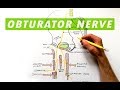 Obturator Nerve - Anatomy Tutorial
 Ultrasound Guided obturator nerve block
 Structures Passing Through Greater Sciatic Notch - Everything You Need To Know - Dr. Nabil Ebraheim
 Sternoclavicular Joint Injuries - Everything You Need To Know - Dr. Nabil Ebraheim
 The obturator artery Animation - Origin , Course , Branches , supply and Clinical anatomy
 Coccydynia - Everything You Need To Know - Dr. Nabil Ebraheim
 Flossing the Obturator Nerve - Amazing Results
 Patient Self Diagnose Carpal Tunnel Syndrome - Everything You Need To Know - Dr. Nabil Ebraheim
 Profunda femoris Artery or Deep femoral artery or The Deep artery of the thigh - Animation
 Nerves of the lower limb
 Superficial Peroneal Nerve Anatomy - Everything You Need To Know - Dr. Nabil Ebraheim
 Ultrasound guided obturator and saphenous nerve block workshop
 Rotator Cuff Muscles - Everything You Need To Know - Dr. Nabil Ebraheim
 Sacral Plexus | Anatomy Tutorial
 Muscles of the Hip and Thigh - Human Anatomy | Kenhub
 Foot Drop, Peroneal Nerve Injury - Everything You Need To Know - Dr. Nabil Ebraheim
 Posterior tibial artery : Origin , Course , Branches , Termination
 Corona Mortis - Everything You Need To Know - Dr. Nabil Ebraheim
 Basics of the Obturator Nerve | Origins & Branches