The obturator artery Animation - Origin , Course , Branches , supply and Clinical anatomy
►𝐉𝐨𝐢𝐧 𝐓𝐡𝐢𝐬 𝐂𝐡𝐚𝐧𝐧𝐞𝐥 𝐓𝐨 𝐆𝐞𝐭 𝐀𝐜𝐜𝐞𝐬𝐬 𝐓𝐨 𝐏𝐞𝐫𝐤𝐬 :- https://bit.ly/2RQHvTN
►𝐃𝐨𝐰𝐧𝐥𝐨𝐚𝐝 𝐭𝐡𝐞 𝐌𝐞𝐝𝐯𝐢𝐳𝐳 𝐚𝐩𝐩 𝐮𝐬𝐢𝐧𝐠 𝐭𝐡𝐞 𝐛𝐞𝐥𝐨𝐰 𝐥𝐢𝐧𝐤 👇👇👇👇 𝐃𝐨𝐰𝐧𝐥𝐨𝐚𝐝 👇👇👇👇
►𝐀𝐧𝐝𝐫𝐨𝐢𝐝 :- https://bit.ly/3ansFKq
📌𝐅𝐨𝐥𝐥𝐨𝐰 𝐨𝐧 𝐈𝐧𝐬𝐭𝐚𝐠𝐫𝐚𝐦 :-
https://www.instagram.com/drgbhanuprakash
The obturator artery Animation - Origin, Course, Branches, supply and Clinical anatomy
The obturator artery is a branch of the anterior division of the internal iliac artery. It provides vascular supply within the pelvis and lower limb.
origin: anterior division of the internal iliac artery
location: pelvis and lower limb
supply: pelvic muscles, ilium, head of femur, muscles of medial thigh, skin
branches: anterior and posterior branches, artery in the ligament of the femur, iliac branch, pubic branch, branch to the knee capsule
The obturator artery most often originates from the anterior division of the internal iliac artery. It travels along the obturator fascia of the pelvic sidewall, between the obturator nerve and vein, to reach the obturator foramen. It is crossed by the ureter close to its origin, and by the ductus deferens in the male.
Branches
---------------
The obturator artery has several branches which may be grouped as follows as branches in the pelvis and those within the thigh.
Branches within the pelvis:
the nutrient artery to the ilium supplies the iliac bone and iliacus muscle
the pubic branch of the obturator artery anastomoses with the pubic branch of the inferior epigastric artery. It then travels through the obturator foramen with the obturator nerve passing into the medial compartment of the thigh.
Within the medial compartment of the thigh the obturator artery splits into anterior and posterior branches. These travel around the origin of the obturator externus and anastomose with one another and the medial circumflex femoral artery.
the anterior branch supplies the adductor brevis muscle, and the skin over the medial thigh at its termination.
the posterior branch gives another branch, the artery of the ligamentum teres which runs in the ligament of the head of the femur and eventually atrophies and becomes nonfunctional after approximately the age of seven. The posterior branch also supplies the adductor muscles and gives a small terminal branch to the capsule of the knee joint.
Supply
----------
The obturator artery supplies the pelvic muscles it crosses, the head of the femur, the muscles of the medial compartment of the thigh and gives a small branch to the knee capsule. The iliac branch supplies the bone and the iliacus muscle. It also has a cutaneous supply to the medial thigh.
#theobturatorartery #obturatorarteryanatomy #anatomyofobturatorartery #obturatorartery #usmle #usmlestep1 #usmleanatomy #obturatorarteryanimation #drgbhanuprakash #drbhanuprakash #uworld #fmge #neetpg
Видео The obturator artery Animation - Origin , Course , Branches , supply and Clinical anatomy канала Dr.G Bhanu Prakash Animated Medical Videos
Показать
Комментарии отсутствуют
Информация о видео
18 декабря 2019 г. 21:12:07
00:01:59
Другие видео канала
 Obturator Artery - Everything You Need To Know - Dr. Nabil Ebraheim
 Obturator nerve Anatomy Animation : Origin, Course , Innervation and Clinical application
 Popliteal Fossa
 Profunda femoris Artery or Deep femoral artery or The Deep artery of the thigh - Animation
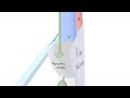 3D Tour of the Femoral Canal
 Functions of the adductor longus muscle (preview) - Human 3D Anatomy | Kenhub
 Obturator Nerve - Anatomy Tutorial
 Pelvic arteries (internal iliac artery)
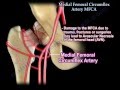 Medial Femoral Circumflex Artery - Everything You Need To Know - Dr. Nabil Ebraheim
 Lower Limb Veins Overview - 3D Anatomy Tutorial
 The Sciatic Nerve Anatomy - Origin, Course, Relations, Branches, Distribution and Clinical anatomy
 Obturator Artery | Anatomy | Branches | Origin and Divisions | Clinical
 Internal Iliac Artery
 The Superior Gluteal Artery - Everything You Need To Know - Dr. Nabil Ebraheim
 Anatomy of Hip Bone / innominate bone / Pelvis ( Osteology ): Ilium, Ischium, Pubis: Animation
 The adductors and the gracilis medial compartment of the thigh
 Great saphenous vein - Animated Gross anatomy of lower limb ( Courtery : Dr vishram singh )
 Radial Nerve | 3D Anatomy Tutorial
 Tibial Nerve Anatomy Animation USMLE Step 1 : Origin, Course, Branches, Tarsal tunnel syndrome
 Anterior and Medial Thigh Anatomy