Obturator Artery - Everything You Need To Know - Dr. Nabil Ebraheim
Dr. Ebraheim’s educational animated video describes anatomy associated with the obturator artery.
The obturator artery is a branch of the anterior division of the internal iliac artery. It arises in the pelvis and it enters the obturator canal.
The obturator artery then divides into two branches:
•Posterior branch of the obturator artery
•Anterior branch of the obturator artery
The anterior and posterior branches of the obturator artery form a vascular circle around the outer surface of the obturator membrane.
The acetabular branch reaches the hip joint and joins the ligamentum teres to supply the head of the femur. It usually supplies a small portion of the head of the femur.
The other branches are muscular branches which supply muscles of the medial side of the thigh.
There are some important anatomical and clinical considerations for the obturator artery.
Corona mortis is a connection between the internal iliac branch (obturator) and the external iliac or its branch, the inferior epigastric. Cadaveric studies show that the incidence of the corona mortis is up to 84%. However, in clinical practice, the incidence of corona mortis is not that high.
Corona mortis is predominantly a venous connection and the arterial connection is much less. Its location on the superior pubic ramus is variable. It is about 3-7 cm from the symphysis pubis. It is located behind and on top of the superior pubic ramus and one must be careful with lateral dissection of the superior pubic ramus.
Corona mortis means the “crown of death”. The corona mortis is susceptible to injury in pelvic trauma and in pelvic surgery. Injury to the corona mortis may lead to significant hemorrhage which may be difficult to control.
Acetabular screws position in total hip arthroplasty. For safe insertion of acetabular screws in total hip arthroplasty, the acetabular anatomy utilizes a quadrant system.
How do you find the safe zone for placement of the acetabular screws? How do you find the danger zone?
A line is drawn from the anterior superior iliac spine through the center of the acetabulum separates the anterior and posterior quadrant. This line is divided or bisected perpendicular to its midpoint to create four quadrants. The obturator vessels are found in the anterior-inferior quadrant. The anterior superior quadrant contains the external iliac vessels.
The obturator artery may be at risk of injury from placement of a retractor underneath the transverse acetabular ligament especially if the retractors are placed too anteriorly during total hip replacement. The transverse acetabular ligament bridges the acetabular notch. The ligament converts the notch into a tunnel. The blood vessels enter the joint through this tunnel. The branches of the obturator artery may bleed in the area of the transverse acetabular ligament. if the transverse acetabular ligament needs to be transacted, then release it in its posterior half, in order to avoid bleeding from the obturator artery.
Reduction of “open book” fracture of the pelvis can be done utilizing reduction clamps. It is probably safe to insert the reduction clamp through the medial side of the foramen, away from the neurovascular bundle which is located laterally.
Follow me on twitter:
https://twitter.com/#!/DrEbraheim_UTMC
Видео Obturator Artery - Everything You Need To Know - Dr. Nabil Ebraheim канала nabil ebraheim
Показать
Комментарии отсутствуют
Информация о видео
Другие видео канала
 Obturator Nerve Anatomy - Everything You Need To Know - Dr. Nabil Ebraheim
 Internal Iliac Artery
 Corona Mortis - Everything You Need To Know - Dr. Nabil Ebraheim
 Femoral artery Anatomy : Origin , Course , Branches and Termination - Animated Lecture
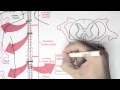 Neurology - Spinal Cord Introduction
 Abdominal aorta - Origin , course , branches - USMLE Step 1 Videos
 "How I Do It: Obturator Bypass" with Dr. Matt Smith and Dr. Sharif Ellozy
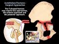 Acetabulum Fractures Surgical Approaches - Everything You Need To Know - Dr. Nabil Ebraheim
 Obturator nerve Anatomy Animation : Origin, Course , Innervation and Clinical application
 Acetabular Fracture Radiology
 MUSCLES OF THE GLUTEAL REGION- ORIGIN, INSERTION, NERVE SUPPLY & ACTION | ANATOMY | SIMPLIFIED ✔
 Obturator Nerve : Only One Page | TCML
 Elbow Pain Causes & Treatment - Everything You Need To Know - Dr. Nabil Ebraheim
 Arteries of the hand
 How to remember the Internal Iliac Artery Branches (The 2+4+4 rule) | Anatomy
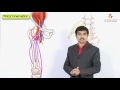 Femoral Nerve Anatomy: Origin, Course, Branches and Clinical application
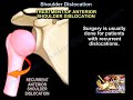 Shoulder Dislocations - Everything You Need To Know - Dr. Nabil Ebraheim
 Pelvic arteries (internal iliac artery)
 Acromioclavicular Joint Injury, AC separation - Everything You Need To Know - Dr. Nabil Ebraheim
 Acetabular Fracture T Shaped Fracture - Everything You Need To Know - Dr. Nabil Ebraheim