Oral Cavity Cancer Staging in 5 minutes
In this video, Dr. Katie Bailey takes on a common topic and describes how cancers of the oral cavity are staged through three quick example cases.
0:00 Introduction
0:24 Review of oral cavity anatomy. The oral cavity includes the lips, teeth, hard and soft palate, gingiva, retromolar trigone, the buccal mucosa, and anterior 2/3 of the tongue.
0:51 Retromolar trigone. The retromolar trigone is a common crossroads where a lot of pathology occurs and it can cause referred ear pain. Mandibular invasion can also occur with masses that occur in this location.
1:34 Oral cavity cancer staging. Tumor (T) staging is based on the size of the tumor and depth of invasion. Nodal (N) staging is based on the number, location, and size of nodes, and metastasis (M) staging is based on the presence or absence of distant sites of disease.
2:21 Example case 1. This example case involves the oral tongue and measures 2-4 cm. It has ipsilateral lymph nodes less than 3 cm. This makes this a T2N2 tumor. Because metastases can’t be evaluated with this information, it is given an ‘X’ for M staging right now.
3:07 Example case 2. This shows a subtle left tongue cancer measuring less than 2 cm. There are no lymph nodes. That makes this a T1N0Mx tumor.
4:01 Example case 3. This case is a hard palate tumor with bone erosion. The involvement through adjacent structures (the bone of the hard palate) makes this a T4 tumor. There is no nodal involvement, making this a T4N0Mx cancer.
Thanks for checking out this quick video on oral cavity cancer staging. Be sure to tune back in for additional videos on staging of the other head and neck subsites.
Check out this video and additional content on http://www.learnneuroradiology.com
Видео Oral Cavity Cancer Staging in 5 minutes канала LearnNeuroradiology
Показать
Комментарии отсутствуют
Информация о видео
Другие видео канала
 Neuroradiology spine lesions - Case 1 - aggressive choice - Choose your own adventure
 Fast 10: Neuroradiology high speed case review part 6 - Cases 51-60
 Neuroradiology board review lecture 1 case 8
 Basic neuroradiology procedures part 3 - Myelogram
 Vascular Imaging of the Head and Neck - Case C
 Neuroradiology Board Review - Brain Tumors - Case 12
 Neuroradiology spine lesions - Case 1 - nonaggressive choice - Choose your own adventure
 Neuroradiology spine lesions - Case 2 - overview - Choose your own adventure
 Nasopharyngeal Cancer Staging in 5 minutes
 Neuroradiology board review 3 case 1
 Neuroradiology board review 3 case 17
 Neuroradiology spine lesions - Case 4 - aggressive choice - Choose your own adventure
 Neuroradiology board review 2 case 10
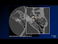 Neuroradiology board review lecture 1 case 15
 Basic Neuroradiology - Chapter 7 - Satisfaction of Search
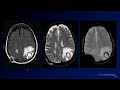 Neuroradiology Board Review - Brain Tumors - Case 3
 Neuroradiology board review 3 case 12
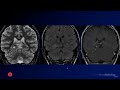 Neuroradiology Board Review - Brain Tumors - Case 16
 Fast 10: Neuroradiology high speed case review part 4 - Cases 31-40
 Neuroradiology board review 3 case 11
 Neuroradiology spine lesions - Choose your own adventure - Introduction