Neuroradiology board review 3 case 11
Neuroradiology board review. This lecture is geared towards the ABR core exam for residents, but it would be useful for review for the ABR certifying exam or certificate of added qualification (CAQ) exam for neuroradiology.
This case shows
a nodular lesion with calcification in the right frontal lobe. There is surrounding vasogenic edema with sparing of the cortex. CT images from higher in the brain show other areas of calcification at the gray-white junction thoughout the brain.
MR confirms these findings. There is an area of right frontal edema with a rounded enhancing structure at the gray-white junction. There is a differential diagnosis for this finding which includes infection and metastatic disease.
The diagnosis is:
neurocysticercosis
Neurocysticercosis is a parasitic infection with a dual life cycle that goes through pigs and humans. When eggs excreted in human feces are ingested, it can affect the CNS. It is the most common cause of acquired seizure in endemic areas.
The treatment is anti-parasitic agents, specifically albendazole.
Check out this video and additional content on http://www.learnneuroradiology.com
Видео Neuroradiology board review 3 case 11 канала LearnNeuroradiology
Показать
Комментарии отсутствуют
Информация о видео
Другие видео канала
 Neuroradiology board review 3 case 12
 NEURORADIOLOGY: CNS Infections - I | DEEPAK PATKAR | Tuberculomas vs Cysticercosis
 Blood brain barrier and vasogenic edema | Circulatory System and Disease | NCLEX-RN | Khan Academy
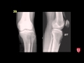 Radiology Boards Prep - MSK Cases
 Your brain on video games | Daphne Bavelier
 Stroke: Acute infarction - radiology video tutorial (CT, MRI, angiography)
 Intracranial infections - 5 - Other
 Neurocisticcircosis symptoms and treatment. Why is pork tapeworm so dangerous?
 Neuroradiology Board Review - Brain Tumors - Case 13
 Neuroradiology Board Review - Brain Tumors - Case 20 - Summary
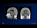 Neuroradiology board review 3 case 1
 Neuroradiology board review 2 case 18
 Cases in Radiology: Episode 1 (neuroradiology, CT, MRI)
 Neuroradiology board review 3 case 10
 Neuroradiology board review 3 case 15
 Neuroradiology Board Review - Brain Tumors - Case 17
 Neuroradiology Board Review - Brain Tumors - Case 19
 Isolation tutorial: Neuroradiology #3 with Frank Gaillard
 Neuroradiology Board Review - Brain Tumors - Case 7
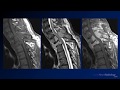 Neuroradiology board review 2 case 10