Neuroradiology board review 3 case 12
Neuroradiology board review. This lecture is geared towards the ABR core exam for residents, but it would be useful for review for the ABR certifying exam or certificate of added qualification (CAQ) exam for neuroradiology.
This case is a patient who has a remote history of "meningitis". Her imaging shows marked edema in the frontal lobes on CT, with a follow up MRI confirming significant leptomeningeal and parenchymal nodular enhancement worst in the frontal lobes. There is involvement of the right optic nerve.
With severe leptomeningeal enhancement such as this, there is a differential diagnosis which includes:
- leptomeningeal carcinomatosis
- unusual infections (fungi, tuberculosis, or other unusual pathogens)
- sarcoidosis
The diagnosis is:
neurosarcoidosis
The most common findings of sarcoidosis are leptomeningeal enhancement centered in the basal cisterns, sometimes with parenchymal nodular or perivascular enhancement. The diagnosis of neurosarcoid consists of checking a serum ACE level, performing an LP (particularly to rule out other causes), and chest imaging (including x-ray or CT). Often the easiest tissue diagnosis is through biopsy of hilar nodes.
Check out this video and additional content on http://www.learnneuroradiology.com
Видео Neuroradiology board review 3 case 12 канала LearnNeuroradiology
Показать
Комментарии отсутствуют
Информация о видео
Другие видео канала
 Neuroradiology spine lesions - Case 1 - aggressive choice - Choose your own adventure
 Fast 10: Neuroradiology high speed case review part 6 - Cases 51-60
 Neuroradiology board review lecture 1 case 8
 Basic neuroradiology procedures part 3 - Myelogram
 Vascular Imaging of the Head and Neck - Case C
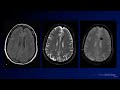 Neuroradiology Board Review - Brain Tumors - Case 12
 Neuroradiology spine lesions - Case 1 - nonaggressive choice - Choose your own adventure
 Neuroradiology spine lesions - Case 2 - overview - Choose your own adventure
 Nasopharyngeal Cancer Staging in 5 minutes
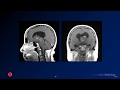 Neuroradiology board review 3 case 1
 Neuroradiology board review 3 case 17
 Neuroradiology spine lesions - Case 4 - aggressive choice - Choose your own adventure
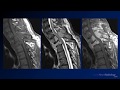 Neuroradiology board review 2 case 10
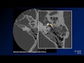 Neuroradiology board review lecture 1 case 15
 Basic Neuroradiology - Chapter 7 - Satisfaction of Search
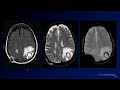 Neuroradiology Board Review - Brain Tumors - Case 3
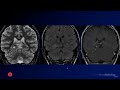 Neuroradiology Board Review - Brain Tumors - Case 16
 Fast 10: Neuroradiology high speed case review part 4 - Cases 31-40
 Neuroradiology board review 3 case 11
 Neuroradiology spine lesions - Choose your own adventure - Introduction