Occipitocervical Dislocation - Everything You Need To Know - Dr. Nabil Ebraheim
Dr. Ebraheim’s educational animated video describes the Occipitocervical Dislocation.
Occipitocervical Dislocation results from disruption of the joint between the skull and the cervical spine. It is a severe injury that cause death to most victims. There will be disruption of all the ligaments and management of the patient is usually difficult. Patients who survive this injury usually have neurological deficit or can have deterioration of their neurological functions. The injury can either occur as:
•Type I: anterior displacement of the occiput.
•Type II: longitudinal distraction of the occiput from the atlas. (Avoid the use of traction).
•Type III: posterior displacement of the occiput.
Radiological interpretation:
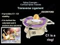
•Basion: it is the mid-point on the anterior margin of the foramen magnum
•Opisthion: it is the mid-point on the posterior margin of the foramen magnum.
Power’s ratio: the normal ratio of BC & AO is 1
Anterior dislocation: if the ratio between BC & AO is greater than 1, an anterior occipitocervical dislocation may exist.
Treatment
•Closed reduction
•Avoid traction in type II, traction is harmful. Traction can cause neurological deficits.
•Occipitocervical fusion is done in most of the cases using plates. Knowledge of the skull anatomy including the thickness of the skull bone and location of the dural sinuses is mandatory. Placement of the screws at this EOP is more likely to injure the dural sinuses.
Become a friend on facebook:
http://www.facebook.com/drebraheim
Follow me on twitter:
https://twitter.com/#!/DrEbraheim_UTMC
Видео Occipitocervical Dislocation - Everything You Need To Know - Dr. Nabil Ebraheim канала nabil ebraheim
Occipitocervical Dislocation results from disruption of the joint between the skull and the cervical spine. It is a severe injury that cause death to most victims. There will be disruption of all the ligaments and management of the patient is usually difficult. Patients who survive this injury usually have neurological deficit or can have deterioration of their neurological functions. The injury can either occur as:
•Type I: anterior displacement of the occiput.
•Type II: longitudinal distraction of the occiput from the atlas. (Avoid the use of traction).
•Type III: posterior displacement of the occiput.
Radiological interpretation:
•Basion: it is the mid-point on the anterior margin of the foramen magnum
•Opisthion: it is the mid-point on the posterior margin of the foramen magnum.
Power’s ratio: the normal ratio of BC & AO is 1
Anterior dislocation: if the ratio between BC & AO is greater than 1, an anterior occipitocervical dislocation may exist.
Treatment
•Closed reduction
•Avoid traction in type II, traction is harmful. Traction can cause neurological deficits.
•Occipitocervical fusion is done in most of the cases using plates. Knowledge of the skull anatomy including the thickness of the skull bone and location of the dural sinuses is mandatory. Placement of the screws at this EOP is more likely to injure the dural sinuses.
Become a friend on facebook:
http://www.facebook.com/drebraheim
Follow me on twitter:
https://twitter.com/#!/DrEbraheim_UTMC
Видео Occipitocervical Dislocation - Everything You Need To Know - Dr. Nabil Ebraheim канала nabil ebraheim
Показать
Комментарии отсутствуют
Информация о видео
Другие видео канала
 Cervical Spine Trauma - Everything You Need To Know - Dr. Nabil Ebraheim
 Muscles of the Back (3D Anatomy Tutorial)
 Brachial Plexus Anatomy Explained - Everything You Need To Know - Dr. Nabil Ebraheim
 Atlas and axis vertebrae
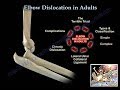 Elbow Dislocation In Adults - Everything You Need To Know - Dr. Nabil Ebraheim
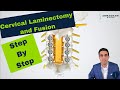 Posterior Cervical Laminectomy and Fusion - Procedure details, recovery, and expectations.
 Spine Emergencies - Everything You Need To Know - Dr. Nabil Ebraheim
 Monteggia Fracture - Everything You Need To Know - Dr. Nabil Ebraheim
 Anterior Shoulder Dislocation
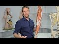 The Patella (anatomy)
 Giant Cell Tumor - Everything You Need To Know - Dr. Nabil Ebraheim
 Disc Herniation - Everything You Need To Know - Dr. Nabil Ebraheim
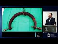 The Dislocated Subaxial C Spine: Reduce First or MRI First - Rick Sasso, MD
 Classifications of Subaxial Cervical Injuries by Andrew T. Dailey, M.D.
 Cervical Flexion & Extension BIOMECHANICS
 Tibial Pilon Fracture - Everything You Need To Know - Dr. Nabil Ebraheim
 Lumbar Spinal Stenosis - Everything You Need To Know - Dr. Nabil Ebraheim
 Cervical Spine X-Ray
 Anatomy Of The Popliteal Fossa - Everything You Need To Know - Dr. Nabil Ebraheim
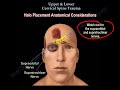 Upper & Lower C Spine Trauma - Everything You Need To Know - Dr. Nabil Ebraheim