Brain imaging course – Unknown case 2
This video is the 2nd unknown case that goes with the brain imaging capstone course.
If you want to follow along, you can find all the images for the case at the brain capstone website:
https://learnneuroradiology.com/braincapstone/
0:00 Introduction
60 year-old man with personality changes and lack of motivation with flat affect for 1-2 months
1:06 Interactive review
In this case, there is an MRI showing a mass in the bilateral frontal lobes, but more in the right frontal lobe. It crosses the corpus callosum. It is markedly enlarged with FLAIR and T2 hyperintensity, abnormal DWI suggesting high cellularity, and a few areas of hemorrhage on SWI.
On post-contrast imaging, you see a mass with peripheral enhancement and central necrosis (a ring enhancing mass). There are multiple additional areas of enhancement (multifocal enhancement). Findings are very concerning for a high grade tumor, such as a glioblastoma.
3:39 Case findings summary
Here you can see screenshots of the findings which we saw in the interactive case review.
4:35 Interactive question
What makes this tumor appear high grade? Central necrosis, thick nodular rind of enhancement, multifocal enhancement, restricted diffusion, crosses midline (corpus callosum)
5:02 Diagnosis and Summary
This is a case of glioblastoma. These are high grade tumors of the brain which have a very poor prognosis, and are one of the few aggressive lesions which will cross from one side of the brain to the other. This is a classic appearance of GBM.
Thanks for tuning in to this case. There are a total of 7 cases you can review on your own at the website and explanations will be posted here.
Check out this video and additional content on http://www.learnneuroradiology.com
Видео Brain imaging course – Unknown case 2 канала LearnNeuroradiology
Показать
Комментарии отсутствуют
Информация о видео
Другие видео канала
 Neuroradiology spine lesions - Case 1 - aggressive choice - Choose your own adventure
 Fast 10: Neuroradiology high speed case review part 6 - Cases 51-60
 Neuroradiology board review lecture 1 case 8
 Basic neuroradiology procedures part 3 - Myelogram
 Vascular Imaging of the Head and Neck - Case C
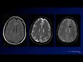 Neuroradiology Board Review - Brain Tumors - Case 12
 Neuroradiology spine lesions - Case 1 - nonaggressive choice - Choose your own adventure
 Neuroradiology spine lesions - Case 2 - overview - Choose your own adventure
 Nasopharyngeal Cancer Staging in 5 minutes
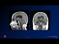 Neuroradiology board review 3 case 1
 Neuroradiology board review 3 case 17
 Neuroradiology spine lesions - Case 4 - aggressive choice - Choose your own adventure
 Neuroradiology board review 2 case 10
 Neuroradiology board review lecture 1 case 15
 Basic Neuroradiology - Chapter 7 - Satisfaction of Search
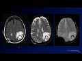 Neuroradiology Board Review - Brain Tumors - Case 3
 Neuroradiology board review 3 case 12
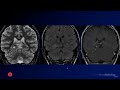 Neuroradiology Board Review - Brain Tumors - Case 16
 Fast 10: Neuroradiology high speed case review part 4 - Cases 31-40
 Neuroradiology board review 3 case 11
 Neuroradiology spine lesions - Choose your own adventure - Introduction