Head and neck anatomy landmarks
Today, Dr. Bailey is back with a video about her approach to head and neck anatomy using landmarks. With this quick video, in about 5 minutes you can learn to quickly differentiate the important anatomical subsites of the head and neck on computed tomography.
0:19 Nasal cavity versus the nasopharynx. The nasal cavity and nasopharynx are both above the hard palate up to the cribriform plate. The nasopharynx begins just behind the posterior margin of the hard palate
1:20 Oral cavity vs oropharynx. Similarly, the oral cavity includes the tissue below the hard palate and anterior to its posterior margin, while the oropharynx includes what is posterior to the margin of the hard palate.
2:30 Floor of the mouth. The floor of the mouth is predominantly made of muscular structures, including the genioglossus, hyoglossus, and mylohyoid.
3:41 Hypopharynx. The hypopharynx consists of the pyriform sinuses, the lateral and posterior pharyngeal walls, and the posterior surfaces of the larynx extending to the cervical esophagus.
4:11 Supraglottic larynx. The supraglottic larynx includes everything from the tip of the epiglottis down to the laryngeal ventricle.
4:45 Larynx-glottis. The glottis includes the larynx and true vocal cords, including the anterior and posterior commissures.
5:17 Larynx-subglottis. The subglottis extends from the inferior aspect of the true vocal cords to the cricoid cartilage. Below the cricoid cartilage is the trachea.
Thanks for tuning in and be sure to come back to check out our additional videos.
Check out this video and additional content on http://www.learnneuroradiology.com
Видео Head and neck anatomy landmarks канала LearnNeuroradiology
Показать
Комментарии отсутствуют
Информация о видео
Другие видео канала
 Neuroradiology spine lesions - Case 1 - aggressive choice - Choose your own adventure
 Fast 10: Neuroradiology high speed case review part 6 - Cases 51-60
 Neuroradiology board review lecture 1 case 8
 Basic neuroradiology procedures part 3 - Myelogram
 Vascular Imaging of the Head and Neck - Case C
 Neuroradiology Board Review - Brain Tumors - Case 12
 Neuroradiology spine lesions - Case 1 - nonaggressive choice - Choose your own adventure
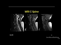 Neuroradiology spine lesions - Case 2 - overview - Choose your own adventure
 Nasopharyngeal Cancer Staging in 5 minutes
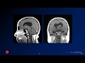 Neuroradiology board review 3 case 1
 Neuroradiology board review 3 case 17
 Neuroradiology spine lesions - Case 4 - aggressive choice - Choose your own adventure
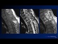 Neuroradiology board review 2 case 10
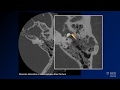 Neuroradiology board review lecture 1 case 15
 Basic Neuroradiology - Chapter 7 - Satisfaction of Search
 Neuroradiology Board Review - Brain Tumors - Case 3
 Neuroradiology board review 3 case 12
 Neuroradiology Board Review - Brain Tumors - Case 16
 Fast 10: Neuroradiology high speed case review part 4 - Cases 31-40
 Neuroradiology board review 3 case 11
 Neuroradiology spine lesions - Choose your own adventure - Introduction