2nd Pharyngeal Arch and its derivatives | Embryology Tutorial
#embryology #pharyngealarch #branchial
Link for Donations https://paypal.me/studentlamedicina?locale.x=en_US
https://www.instagram.com/anatomy.knowledge/
The second pharyngeal arch , also called The hyoid arch, appears during the 4th week of development and is situated distally to the first pharyngeal arch and laterally to the primitive pharynx.
On a cross section the second paryngeal arch presents the typical features of a pharyngeal arch.
A core of mesoderm covered internally by endoderm and externally by ectoderm. A cartilage, a nerve, and an aortic arch.
In the structure of the second pharyngeal arch is the Reichert’s Cartilage. This cartilage contributes to the formation of the following structures:
Lesser cornu and upper part of the body of the hyoid bone,
The Styloid process of the temporal bone,
Stylohyoid ligament,
And Stapes.
The nerve of the second pharyngeal arch is the facial nerve.
The facial nerve innervates all the following muscles derived from the second pharyngeal arch:
Occipitofrontalis muscle,
Platysma muscle,
Facial muscles,
Auricular muscles,
Stylohyoid ,
Posterior belly of digastric,
And the stapedius muscle.
The artery of the second pharyngeal arch is the second aortic arch, which gives origin to the hyoid artery and its branch, the stapedial artery. The stapedial artery atrophies while remnants of the hyoid artery persist in adults as the caroticotympanic arteries.
Видео 2nd Pharyngeal Arch and its derivatives | Embryology Tutorial канала Anatomy Knowledge
Показать
Комментарии отсутствуют
Информация о видео
Другие видео канала
 Pharyngeal Arches and its Derivatives - MASTER pharyngeal arches in LESS than 7 minutes ONLY!
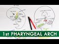 First Pharyngeal Arch and its derivatives | Embryology Tutorial
 Development of Tongue - (Embryology video)
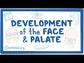 Development of the Face and Palate
 Pharyngeal arches | Embryology
 Forearm Muscles - Anterior Compartment | Anatomy Tutorial
 Muscles of the Arm - Origin, Insertion, Innervation | Anatomy Tutorial
 Embryology of the Pharyngeal Arches (Easy to Understand)
 Muscles of the neck
 Muscles of Mastication | Anatomy Tutorial
 Posterior (Extensor) Compartment of Forearm | Anatomy Tutorial
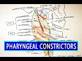 Pharyngeal Constrictors | Pharynx Anatomy
 General Embryology Review in 20 minutes
 Pharyngeal Arches Mnemonic | Embryology | Pharyngeal Arch Derivatives Mnemonic Anatomy
 Cleft Lip and Palate - Pathophysiology, Causes & Management
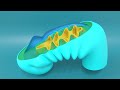 3D Embryology of Pharyngeal arches, Pharyngeal Pouches, Pharyngeal clefts and Pharyngeal Apparatus
 Development of thyroid gland- Chart | TCML
 Pharyngeal arches and their derivatives
 FOURTH VENTRICLE | NEUROANATOMY | SIMPLIFIED
 Anterior Cranial Fossa | Anatomy Tutorial