Posterior (Extensor) Compartment of Forearm | Anatomy Tutorial
#extensorcompartment #forearm #radial
Link for Donations https://paypal.me/studentlamedicina?locale.x=en_US
https://www.instagram.com/anatomy.knowledge/
There are twelve muscles in the posterior compartment of forearm which can be further divided into three groups.
First group to be described is the lateral group which contains three muscles.
The brachioradialis arises from the upper two-third of the lateral supracondylar ridge
of the humerus. Its tendon inserts into the lateral surface of the distal end of radius just above the styloid process.
Having origin from the lower one-third of the lateral supracondylar ridge of the humerus and inserting into the base of the second metacarpal bone is the extensor carpi radialis longus muscle.
Having origin from the lateral epicondyle of humerus and inserting into the base of the third metacarpal bone is the extensor carpi radialis brevis muscle.
The next group of muscles to be described is the posterior superficial group which contains four muscles.
Having origin from the lateral epicondyle of humerus and inserting into the lateral side of the olecranon process and upper one fourth of the posterior surface of the ulna is the anconeus muscle.
Having origin from the lateral epicondyle of humerus and inserting into the medial side of the base of the fifth metacarpal bone is the extensor carpi ulnaris.
The biggest muscle of the posterior superficial group is the extensor digitorum. This muscle arises from the lateral epicondyle of humerus and gives rise to four tendons for medial four digits. Each tendon gives three slips. The middle slip inserts into the base of the second phalanx of the cresponding finger. The lateral two tendinous slips inserts into the base of the distal phalanx of the coresponding finger.
Lying medially to extensor digitorum is a small muscle called the extensor digiti minimi. This muscle also originates from the lateral epicondyle of humerus. Distaly, its tendon fuses with the extensor digitorum tendon for little finger.
The last group of muscles in the posterior compartment of forearm is the posterior deep group. This group contains five muscle.
First muscle to be indicated is the supinator muscle. This muscle arises from the lateral epicondyle of humerus, supinator crest of ulna, radial collateral ligament and annular ligament. Its insertion is into the upper one-third of the lateral surface of radius.
The next muscle to be indicated is the abductor policis longus. The origin of this muscle is from the posterior surfaces of ulna, radius and interosseous membrane. The tendon of abductor policis longus inserts into the base of the first metacarpal bone.
Having origin from a small area on the posterior surface of radius below the origin of abductor pollicis longus and from adjoining interosseous membrane is the extensor policis brevis. Its tendon inserts into the dorsal surface of the base of the proximal phalanx of the thumb.
Having origin from lateral part of middle one-third of the posterior surface of ulna and adjoining interosseous membrane is the extensor policis longus muscle. Its tendon inserts into the base of the distal phalanx of thumb.
The last muscle to be indicated is the extensor indicis. This muscle arises from the posterior
surface of ulna below the origin of extensor pollicis longus and also from the adjoining interosseous membrane. Its tendon fuses whith the extensor digitorum tendon from the index finger.
Видео Posterior (Extensor) Compartment of Forearm | Anatomy Tutorial канала Anatomy Knowledge
Показать
Комментарии отсутствуют
Информация о видео
Другие видео канала
 Forearm Muscles - Anterior Compartment | Anatomy Tutorial
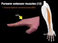 Forearm extensor muscles
 Musculocutaneous Nerve - Anatomy Diagram
 Forearm Muscles Part 2 - Posterior (Extensor) Compartment - Anatomy Tutorial
 How To Remember Every Muscle in the Upper Limb and Arm | Corporis
 Clinical Anatomy - Hand, Wrist (palmar aspect/flexors)
 Posterior compartment of forearm muscles (preview) - Human Anatomy | Kenhub
 Superficial Veins of Upper Limb - Basilic & Cephalic veins | Anatomy Tutorial
 Muscles of the Hand - Origin, Insertion, Nerve Supply | Anatomy Tutorial
 Posterior forearm muscles (identifying)
 Median Nerve | Anatomy Tutorial
 Radial Nerve Anatomy - Everything You Need To Know - Dr. Nabil Ebraheim
 Anatomy of the Forearm - Muscles and Tendons - Lesson 1
 Muscles of Shoulder - Origins, Insertions, Innervations | Anatomy Tutorial
 Muscles of the Anterior Forearm - Animated Anatomy
 Axillary Nerve - Course & Branches | Anatomy Tutorial
 Median Nerve | 3D Anatomy Tutorial
 Lockdown Anatomy with Prof Alice Roberts #4: forearm flexors
 Muscles of the Arm - Origin, Insertion, Innervation | Anatomy Tutorial
 Hand Muscles