Necrosis and types of Necrosis , General pathology - Animated usmle videos
►𝐉𝐨𝐢𝐧 𝐓𝐡𝐢𝐬 𝐂𝐡𝐚𝐧𝐧𝐞𝐥 𝐓𝐨 𝐆𝐞𝐭 𝐀𝐜𝐜𝐞𝐬𝐬 𝐓𝐨 𝐏𝐞𝐫𝐤𝐬 :- https://bit.ly/2RQHvTN
►𝐃𝐨𝐰𝐧𝐥𝐨𝐚𝐝 𝐭𝐡𝐞 𝐌𝐞𝐝𝐯𝐢𝐳𝐳 𝐚𝐩𝐩 𝐮𝐬𝐢𝐧𝐠 𝐭𝐡𝐞 𝐛𝐞𝐥𝐨𝐰 𝐥𝐢𝐧𝐤 👇👇👇👇 𝐃𝐨𝐰𝐧𝐥𝐨𝐚𝐝 👇👇👇👇
►𝐀𝐧𝐝𝐫𝐨𝐢𝐝 :- https://bit.ly/3ansFKq
📌𝐅𝐨𝐥𝐥𝐨𝐰 𝐨𝐧 𝐈𝐧𝐬𝐭𝐚𝐠𝐫𝐚𝐦 :-
https://www.instagram.com/drgbhanuprakash
Necrosis and types of Necrosis , General pathology - Animated usmle videos
Coagulative
-------------------
See this in infarcts in any tissue (except brain)
Due to loss of blood
Gross: tissue is firm
Micro: Cell outlines are preserved (cells look ghostly), and everything looks red
Liquefactive
--------------------
See this in infections and, for some unknown reason, in brain infarcts
Due to lots of neutrophils around releasing their toxic contents, “liquefying” the tissue
Gross: tissue is liquidy and creamy yellow (pus)
Micro: lots of neutrophils and cell debris
Caseous
--------------
See this in tuberculosis
Due to the body trying to wall off and kill the bug with macrophages
Gross: White, soft, cheesy-looking (“caseous”) material
Micro: fragmented cells and debris surrounded by a collar of lymphocytes and macrophages (granuloma)
Fat necrosis
--------------------
See this in acute pancreatitis
Damaged cells release lipases, which split the triglyceride esters within fat cells
Gross: chalky, white areas from the combination of the newly-formed free fatty acids with calcium (saponification)
Micro: shadowy outlines of dead fat cells (see image above); sometimes there is a bluish cast from the calcium deposits, which are basophilic
Fibrinoid necrosis
-----------------------------
See this in immune reactions in vessels
Immune complexes (antigen-antibody complexes) and fibrin are deposited in vessel walls
Gross: changes too small to see grossly
Micro: vessel walls are thickened and pinkish-red (called “fibrinoid” because the deposits look like fibrin deposits)
Gangrenous necrosis
-----------------------------------
See this when an entire limb loses blood supply and dies (usually the lower leg)
This isn’t really a different kind of necrosis, but people use the term clinically so it’s worth knowing about
Gross: skin looks black and dead; underlying tissue is in varying stages of decomposition
Micro: initially there is coagulative necrosis from the loss of blood supply (this stage is called “dry gangrene”); if bacterial infection is superimposed, there is liquefactive necrosis (this stage is called “wet gangrene”)
#necrosis #pathovideos #usmlevideos #necrosispathology #pathologyofnecrosis #usmlestep1videos #animatedmedicalvideos #pathoanimatedvideos #drgbhanuprakash #mbbs #fmge #neetpg
Видео Necrosis and types of Necrosis , General pathology - Animated usmle videos канала Dr.G Bhanu Prakash Animated Medical Videos
Показать
Комментарии отсутствуют
Информация о видео
8 декабря 2017 г. 16:04:57
00:10:22
Другие видео канала
 Necrosis - Cell Injury - General Pathology
 What is Necrosis vs What is Apoptosis?
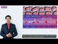 1.Edema introduction - Hemodynamic pathology Fmge, Neet PG and usmle
 Apoptosis (Intrinsic, Extrinsic Pathways) vs. Necrosis
 PATHOLOGIC CALCIFICATION: Dystrophic & Metastatic Calcification
 AMYLOIDOSIS: Part 1: Definition, Historical aspects & Properties of Amyloid
 Immunology | Inflammation: Vascular Events | Part 1
 Cell Adaptations: pathology:HYPERTROPHY HYPERPLASIA AND METAPLASIA
 Physiology of wound healing
 Osteoporosis - causes, symptoms, diagnosis, treatment, pathology
 Necrosis & Types of Necrosis Simplified
 Gangrene: Dry, Wet and Gas Gangrene
 Necrosis | Necrosis Types | Necrosis Patterns | Necrosis animation | Necrosis Pathology Lecture |
 Sadhguru - How can you fight cancer ?!
 TISSUR REPAIR Part 3: WOUND HEALING, Factors affecting wound healing.
 Necrosis : cellular mechanism and types
 APOPTOSIS PART 1: Definition, Causes & Mechanism/Pathways
 Cell Injury ( Part 2 ) : Various Mechanisms, Reversible & Irreversible Injury, Necrosis (HD)
 Inflammation - Vascular Events
 MORPHOLOGY OF CELL INJURY: REVERSIBLE & IRREVERSIBLE. Necrosis & its types.