Trochlear Nerve | Anatomy Tutorial
#trochlear #cranialnerve #anatomy
Link for Donations https://paypal.me/studentlamedicina?locale.x=en_US
https://www.instagram.com/anatomy.knowledge/
The trochlear nerve is the smallest of the cranial nerves and innervates a single muscle in the orbit, the superior oblique muscle.
The axons of the trochlear nerve originate in the trochlear nucleus located in the tegmentum of the midbrain at the level of the inferior colliculus, and ventro lateral to the periaqueductal gray matter.
Axons arising from the trochlear nucleus course dorsally around the periaqueductal grey matter and cross the midline. The crossed axons emerge from the dorsal aspect of the midbrain just caudal to the inferior colliculus to form cranial nerve IV or the trochlear nerve.
The nerve curves ventrally around the cerebral peduncle to pass between the posterior cerebral and superior cerebellar arteries.
The trochlear nerve enters the lateral wall of cavernous sinus where is situated between oculomotor and ophthalmic nerves.
It leaves the cavernous sinus and enters the orbit through the superior orbital fissure, above
the tendinous ring. The nerve then courses medially, close to the roof of the orbit, and runs diagonally above the levator palpebrae superioris muscle to reach its target, the superior oblique muscle. Here the nerve divides into three or more branches that enter the superior oblique muscle along its proximal third.
The trochlear nerve is unique among the cranial nerves in several respects:
• It is the smallest nerve in terms of the number of axons it contains (approximately 2400 axons).
• It has the greatest intracranial length.
• It is the only cranial nerve that exits from the dorsal aspect of the brainstem.
• It is the only cranial nerve whose nuclear fibres decussate
before emerging on the surface of the brain.
• Its nucleus receives only ipsilateral corticonuclear fibres.
Видео Trochlear Nerve | Anatomy Tutorial канала Anatomy Knowledge
Показать
Комментарии отсутствуют
Информация о видео
Другие видео канала
 Inferior surface of the brain - Lobes, Gyri, Sulci | Neuroanatomy
 Medial surface of the Cerebral hemisphere - Gyri and sulci | Neuroanatomy
 Scalene Muscles | Scalene Hiatus - Anatomy Tutorial
 Abducens Nerve - Anatomy Tutorial
 The Subinguinal Space - Lacuna Vasorum & Lacuna Musculorum | Anatomy Tutorial
 Anterior view of the brainstem | Neuroanatomy Diagram
 Thigh cross sectional anatomy - Muscular Compartments - Adductor Canal
 Popliteal Fossa | Boundaries & Contents | Anatomy Tutorial
 Gluteal region | Anatomy Tutorial - Greater sciatic foramen & lesser sciatic foramen
 Anatomy - Lumbar Plexus
 Venous drainage of Stomach | Anatomy Tutorial
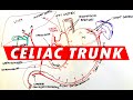 Celiac Trunk - Arterial supply to the Stomach
 Thoracolumbar Fascia | Anatomy Tutorial
 Hypoglossal Nerve | Course & Branches | Anatomy Tutorial
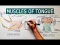 Muscles of the Tongue | Anatomy tutorial
 Anatomy Tutorial - Superior Thyroid Artery
 BRACHIAL PLEXUS - Anatomy Tutorial
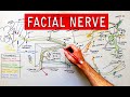 Facial nerve - Origin, Function, Pathway & Branches | Anatomy Tutorial
 The Infratemporal Fossa - Boundaries & Contents | Anatomy Tutorial
 Radial Nerve - part #1 | Anatomy Tutorial
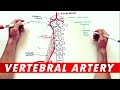 Anatomy Tutorial - The Vertebral Artery