Facial nerve - Origin, Function, Pathway & Branches | Anatomy Tutorial
#facialnerve #cranial #trigeminal
Donation Link: https://paypal.me/studentlamedicina?locale.x=en_US
https://www.instagram.com/anatomy.knowledge/
Facial nerve is the 7th cranial nerve. It is a mixed nerve, but predominantly it is motor.
Four nuclei are related to the fibers which travel within the facial nerve:
- Motor nucleus of facial nerve located in pons;
- Superior salivatory nucleus;
- Spinal nucleus of trigeminal nerve;
- Nucleus solitarius.
Upon emerging from the stylomastoid foramen, the facial nerve gives rise to the posterior auricular nerve. This branch runs upward in front of the mastoid process and splits into two branches: an auricular branch which supplies the posterior auricular muscle and an occipital branch which supplies the occipitalis muscle.
Another two small branches are arising from the facial nerve soon after emerging from the stylomastoid foramen. Branch to stylohyoid muscle and branch to posterior belly of digastric muscle.
Within the substance of the parotid gland five major branches are arising from the facial nerve which will innervate all the muscles of facial expression. These branches , from top to bottom are as follows:
The temporal branch
The zygomatic branch
The buccal branch
The marginal mandibular branch
The Cervical branch.
From these branches the most important one is the zygomatic branch which supplies the orbicularis occuli muscle. This muscle closes the eyelids.
An injury to the zygomatic branch would alter the function of the ipsilateral oribularis occuli muscle, the eyelids would be unable to close thus inflammation, dryness, ulceration or even blindness may occur in extreme cases.
Видео Facial nerve - Origin, Function, Pathway & Branches | Anatomy Tutorial канала Anatomy Knowledge
Показать
Комментарии отсутствуют
Информация о видео
Другие видео канала
 Neurology | Facial Nerve: Cranial Nerve VII
 Trigeminal Nerve Anatomy - The Ophthalmic Nerve
 The Anterior Triangle of the Neck - Boundaries ❌ Subdivisions | Anatomy Tutorial
 Gross Anatomy of the Middle Ear - Boundaries ,Contents and Functions ( Animation )
 Internal Jugular Vein | Anatomy Tutorial
 Lesser Omentum - Attachment, Ligaments & Contents | Anatomy Tutorial
 Pons | Cross section | Internal structure - Neuroanatomy Tutorial
 MAXILLARY ARTERY and its Branches - Anatomy Tutorial
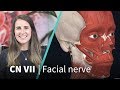 Anatomy Dissected: Cranial Nerve VII (facial nerve)
 The Posterior Triangle of the Neck - Boundaries & Content - Head & Neck Anatomy
 Cervical Plexus - Anatomy Tutorial
 Pterygopalatine Fossa - Boundaries, Communications & Contents
 Facial Nerve - Neuroanatomy - Part 1/4
 Glossopharyngeal Nerve | Cranial Nerve IX | Anatomy Tutorial
 Vagus Nerve | Cranial nerve X - Head & Neck Anatomy Tutorial
 Cranial Nerve BASICS - The 12 cranial nerves and how to REMEMBER them!
 The Facial Nerve (CNVII): Animated Review
 Ciliary Ganglion - Autonomic control of the eye | Anatomy Tutorial
 Cranial Nerves Exam | Clinical Skills
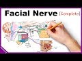 Facial Nerve Anatomy Simplified