The Posterior Triangle of the Neck - Boundaries & Content - Head & Neck Anatomy
Link to PayPal donation https://paypal.me/studentlamedicina?locale.x=en_US
https://www.instagram.com/anatomy.knowledge/
The posterior triangle of the neck is the triangular space on the side of neck behind the sternocleidomastoid muscle. Its apex is directed upwards and backwards towards the mastoid process and base downwards towards the clavicle.
Boundaries:
Anterior is bounded by the posterior border of sternocleoidomastoid muscle.
Posterior is the anterior margin of trapezius muscle.
Inferiorly is bounded by the superior aspect of middle third of the clavicle.
The floor of posterior triangle is muscular and is formed from above downwards by the following muscles:
1. Semispinalis capitis.
2. Splenius capitis.
3. Levator scapulae.
4. Scalenus medius.
5.and on a smal area the Scalenus anterior is also observed. Most of scalenus anterior lies behind the inferior extremity of sternocleidomastoid muscle.
Through the scalene hiatus passes the subclavian artery and trunks of brachial plexus. In front of the scalenus anterior passes the phrenic nerve which is oriented vertically, and the subclavian vein which is oriented horizontally. At this level the subclavian vein recieves the external jugular vein.
#anatomy #head #neck
Видео The Posterior Triangle of the Neck - Boundaries & Content - Head & Neck Anatomy канала Anatomy Knowledge
Показать
Комментарии отсутствуют
Информация о видео
Другие видео канала
 The Anterior Triangle of the Neck - Boundaries ❌ Subdivisions | Anatomy Tutorial
 Suprahyoid & Infrahyoid muscles of the neck | Anatomy Tutorial
 Carotid triangle - boundaries & contents | Anatomy Tutorial
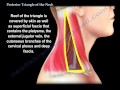 Posterior Triangle Of The Neck - Everything You Need To Know - Dr. Nabil Ebraheim
 Posterior Triangle Of the Neck | Boundaries | Subdivisions | Contents
 Carotid triangle - Animated Gross anatomy head and neck , medical animation
 ANATOMY Tutorial - External Carotid Artery Branches
 Muscles of the Tongue | Anatomy tutorial
 Posterior Triangle of Neck ( Boundary , floor )
 Arterial supply of the Thorax - Anatomy Tutorial
 Facial nerve - Origin, Function, Pathway & Branches | Anatomy Tutorial
 Submandibular triangle - boundaries & contents | Anatomy Tutorial
 Triangles of neck made easy!
 Cervical fascia
 Neck: Cervical Compartments & Cervical Fascia – Brain & Nervous System | Lecturio
 TRIANGLES OF NECK
 Triangles of the Neck
 The Suboccipital Triangle | Boundaries | Contents
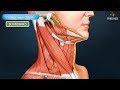 Digastric triangle : Boundaries and contents - Animated Gross anatomy head and neck
 Triangles of Neck || SIMPLIFIED || Dr. Yusuf ||