Carotid triangle - Animated Gross anatomy head and neck , medical animation
FOLLOW ON INSTAGRAM :- https://www.instagram.com/drgbhanuprakash/
Channel Memberships : https://www.youtube.com/channel/UCG5TBPANNSiKf1Dp-R5Dibg/join
Carotid triangle
-------------------------
The carotid triangle is one of the paired triangles in the anterior triangle of the neck. The triangles of the neck are surgically focussed, first described from early dissection-based anatomical studies which predated cross-sectional anatomical description based on imaging (see deep spaces of the neck).
Boundaries
-------------------
superior: posterior belly of digastric
anteroinferiorly: superior belly of omohyoid
posterior: sternocleidomastoid
floor: thyrohyoid, hyoglossus, middle and inferior pharyngeal constrictors
roof: skin, superficial fascia, platysma, deep fascia
Contents
---------------
Important vascular structures are found within this triangle. It contains the bifurcation of the common carotid artery into internal and external carotid arteries.
The internal carotid artery initially lies postero-laterally to the external carotid artery. It then becomes posterior to it.
Branches of the external carotid artery are found in the triangle including:
superior thyroid artery
lingual artery
facial artery
occipital artery
ascending pharyngeal artery
Corresponding veins to these branches drain into the internal jugular vein.
Crossing both the internal and external carotid artery is the hypoglossal nerve.
Nerves
-----------
hypoglossal nerve
nerve to thyrohyoid from the C1 ventral ramus of the cervical plexus
superior root of ansa cervicalis
internal laryngeal nerve
external laryngeal nerve
#carotidtriangle #anatomyofcarotidtriangle #usmlevideos #carotidtrangleanatomy #animatedmedicalvideos #animatedanatomyvideos #usmlestep1videos #usmleanatomyvideos #mbbs #bhanuprakashanatomyvideos #neetpg #fmge
Видео Carotid triangle - Animated Gross anatomy head and neck , medical animation канала Dr.G Bhanu Prakash Animated Medical Videos
Показать
Комментарии отсутствуют
Информация о видео
30 октября 2017 г. 17:45:18
00:03:33
Другие видео канала
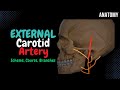 External Carotid Artery (Side branches + Mnemonics)
 ANSA CERVICALIS - USMLE STEP 1 : ANIMATED ANATOMY VIDEO LECTURE
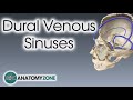 Dural Venous Sinuses | 3D Anatomy Tutorial
 Anatomy and Physiology of Larynx , Action of Laryngeal muscles , Dr Bhanu prakash
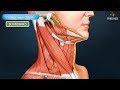 Digastric triangle : Boundaries and contents - Animated Gross anatomy head and neck
 Triangles of the Neck
 Anatomy of Temporomandibular joint ( TMJ ) Head and Neck - Gross Anatomy medical animations
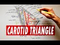 Carotid triangle - boundaries & contents | Anatomy Tutorial
 The Sciatic Nerve Anatomy - Origin, Course, Relations, Branches, Distribution and Clinical anatomy
 Radial nerve Anatomy - Origin, Course, innervation, Saturday night palsy, Wartenberg’s syndrome
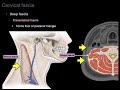 Cervical fascia
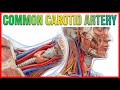 Common carotid Artery Anatomy - Origin , Course , Relations , Branches , Clinical anatomy - USMLE
 Coronary arteries Anatomy / Blood supply of Heart / Arterial supply of heart : Animation
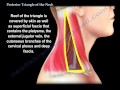 Posterior Triangle Of The Neck - Everything You Need To Know - Dr. Nabil Ebraheim
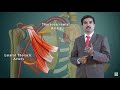 AXILLARY ARTERY ANATOMY ANIMATED LECTURE
 The Posterior Triangle of the Neck - Boundaries & Content - Head & Neck Anatomy
 THYROID GLAND | ANATOMY | SIMPLIFIED
 Muscles of the neck
 CAROTID TRIANGLE
 Inner ear Anatomy : Cochlear component, Vestibular component, Semi-circular component - Animation