Vagus Nerve | Cranial nerve X - Head & Neck Anatomy Tutorial
#vagus #anatomy #larynx
Link for Donations https://paypal.me/studentlamedicina?locale.x=en_US
https://www.instagram.com/anatomy.knowledge/
The vagus nerve is the 10th cranial nerve and is a mixed nerve. Its field of distribution extends beyond the head and neck—to the thorax and abdomen, but in our tutorial we will focus on its distribution in the head and neck region.
The vagus nerve arises from the lateral aspect of the medulla between the olive and inferior cerebellar peduncle and exits the skull through the jugular foramen.
Its two sensory ganglia, the superior (jugular) and the inferior
(nodosum) are located on the nerve.
As the vagus continues below the inferior ganglion, it is joined by the motor fibers from the nucleus ambiguus that have travelled briefly with the cranial root of accessory nerve.
The vagus have four related nuclei located in the medulla. The nucleus ambiguus, the dorsal nucleus of vagus, the nucleus solitarius and the spinal nucleus of trigeminal nerve.
Its Meningeal branch arises from the superior ganglion, takes a recurrent course, and enters the cranial cavity through the jugular foramen to supply the dura mater of the posterior cranial fossa.
The Auricular branch or the Arnold’s nerve arises from the superior ganglion, enters the mastoid canaliculus on the lateral wall of the jugular fossa, and emerges through the tympanomastoid fissure just behind the external auditory meatus to supply the skin on the back of the meatus and adjoining part of the auricle, the floor of the meatus and the tympanic membrane.
The pharyngeal branch, the principal motor nerve of the pharynx, traverses the inferior ganglion and passes inferomedially to enter the pharynx at the upper border of the middle constrictor and breaks up into the pharyngeal plexus to supply all of the muscles of the pharynx and soft palate except stylopharyngeus and tensor palati.
The superior laryngeal nerve arises from the inferior ganglion or just bellow it and passes downward and forward to reach the middle constrictor where it divides into internal and external laryngeal nerves. The internal laryngeal nerve is a sensory nerve and pierces the thyrohyoid membrane to supply the mucous membrane of the pharynx, epiglottis, vallecula, and the posteriormost part of the tongue and also the mucous membrane of the larynx above the vocal cords. The external laryngeal nerve is motor and runs downward to supply the cricothyroid muscle and part of the inferior constrictor.
We have two cervical cardiac branches from the vagus nerve. The superior cardiac branch arises in the upper part of the neck and the inferior cardiac branch which arises in the lower part of the neck.
They enter the thorax through the thoracic inlet and carry preganglionic parasympathetic cardio-inhibitory fibres to the heart.
We have one more branch to indicate from the vagus nerve in the head and neck region and this is the recurrent laryngeal nerve.
There are two recurrent laryngeal nerves, right and left and are not symmetrical, with the left nerve looping under the aortic arch, and the right nerve looping under the right subclavian artery then traveling upwards to reach the tracheoesophageal groove. Each recurrent laryngeal nerve passes deep to the inferior constrictor muscle to enter the laryngeal cavity just posterior to the cricothyroid joint.
The recurrent laryngeal nerve is a mixed nerve and provides motor innervation to all the intrinsic muscles of the larynx except the cricothyroid and sensory innervations to the mucous membrane of laryngeal cavity bellow the vocal cords.
Видео Vagus Nerve | Cranial nerve X - Head & Neck Anatomy Tutorial канала Anatomy Knowledge
Показать
Комментарии отсутствуют
Информация о видео
Другие видео канала
 Cranial Nerve BASICS - The 12 cranial nerves and how to REMEMBER them!
 Glossopharyngeal Nerve | Cranial Nerve IX | Anatomy Tutorial
 Five Tips for Boosting your Vagus Nerve
 Facial nerve - Origin, Function, Pathway & Branches | Anatomy Tutorial
 What Is The Vagus Nerve? | Vagus Nerve Explained | Brain, Mind Body Connect
 Anatomy Dissected: Cranial Nerve V (trigeminal nerve)
 The Facial Nerve (CNVII): Animated Review
 Hypoglossal Nerve | Course & Branches | Anatomy Tutorial
 Gastrointestinal (GI) symptoms and the vagus nerve - cervical instability can cause GI dysfunction
 Cervical Plexus - Anatomy Tutorial
 Superficial Veins of Upper Limb - Basilic & Cephalic veins | Anatomy Tutorial
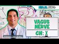 Neurology | Vagus Nerve: Cranial Nerve X
 Cervical Dysstructure and digestive problems - the vagus nerve connection
 Anatomy - Cranial Nerves Overview
 Cervical fascia
 Facial Nerve Anatomy Simplified
 Vagus Nerve & Accessory Nerve | Course and Distribution
 Neurology | Autonomic Nervous System
![Cranial Nerve X - Vagus Nerve [Part 2a] | Structure & Functions of UQ & Thoracic Branches](https://i.ytimg.com/vi/KtWAQxkINNo/default.jpg) Cranial Nerve X - Vagus Nerve [Part 2a] | Structure & Functions of UQ & Thoracic Branches
Cranial Nerve X - Vagus Nerve [Part 2a] | Structure & Functions of UQ & Thoracic Branches Vagus Nerve Massage For Stress And Anxiety Relief