Gross Anatomy of the Middle Ear - Boundaries ,Contents and Functions ( Animation )
Follow On Instagram :- https://www.instagram.com/drgbhanuprakash
Channel Memberships : https://www.youtube.com/channel/UCG5TBPANNSiKf1Dp-R5Dibg/join
Gross anatomy of the Middle Ear - Boundaries, Contents and Functions (Animation)
The ear can be split into three parts; external, middle and inner.
The middle ear lies within the temporal bone, and extends from the tympanic membrane to the lateral wall of the inner ear. The main function of the middle ear is to transmit vibrations from the tympanic membrane to the inner ear via the auditory ossicles.
Parts of the Middle Ear
The middle ear can be divided into two parts:
Tympanic cavity – located medially to the tympanic membrane. It contains three small bones known as the auditory ossicles: the malleus, incus and stapes. They transmit sound vibrations through the middle ear.
Epitympanic recess – a space superior to the tympanic cavity, which lies next to the mastoid air cells. The malleus and incus partially extend upwards into the epitympanic recess.
Borders
The middle ear can be visualised as a rectangular box, with a roof and floor, medial and lateral walls and anterior and posterior walls.
Roof – formed by a thin bone from the petrous part of the temporal bone. It separates the middle ear from the middle cranial fossa.
Floor – known as the jugular wall, it consists of a thin layer of bone, which separates the middle ear from the internal jugular vein
Lateral wall – made up of the tympanic membrane and the lateral wall of the epitympanic recess.
Medial wall – formed by the lateral wall of the internal ear. It contains a prominent bulge, produced by the facial nerve as it travels nearby.
Anterior wall – a thin bony plate with two openings; for the auditory tube and the tensor tympani muscle. It separates the middle ear from the internal carotid artery.
Posterior wall (mastoid wall) – it consists of a bony partition between the tympanic cavity and the mastoid air cells.
Superiorly, there is a hole in this partition, allowing the two areas to communicate. This hole is known as the aditus to the mastoid antrum.
#middleearanatomyanimation #middleear #drgbhanuprakash #middleeargrossanatomy #anatomyofmiddleear #middleearboundaries #middleearcontents #middleearfunctions #middleeargrossanatomy #middleearvideo #middleearusmle #middleearexplained #middleearlecture #middleearanatomy #earanatomy #anatomyoftheear #middleearfunction #middleearborders #middleearanimation #anatomyoftheear #usmlestep1 #earanatomy
Видео Gross Anatomy of the Middle Ear - Boundaries ,Contents and Functions ( Animation ) канала Dr.G Bhanu Prakash Animated Medical Videos
Показать
Комментарии отсутствуют
Информация о видео
3 января 2020 г. 18:11:28
00:13:43
Другие видео канала
 Inner ear Anatomy : Cochlear component, Vestibular component, Semi-circular component - Animation
 Tympanic Membrane Anatomy - Head and neck , Medvizz Anatomy medical animations : Usmle step 1
 Special Senses | External & Middle Ear Anatomy
 Inflammatory and infectious disease of the temporal bone Harn
 MASTOID ANTRUM and AIR CELLS complete explanation in easy way
 Anatomy of the Human Ear
 NEUROANATOMY-THE BRAINSTEM-PART 1 THE MEDULLA OBLONGATA-DR ROSE JOSE MD
 Human ear - structure & working | Sound | Physics | Khan Academy
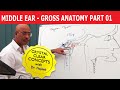 Middle Ear - Gross Anatomy - Part 1/9
 middle ear mucosal folds and spaces
 Gross anatomy of lower limb - FRONT OF THE THIGH PART 2
 Middle ear (tympanic cavity) anatomy
 ANATOMY OF EXTERNAL EAR - Dr.G.Bhanu Prakash
 Middle Ear Anatomy - 1
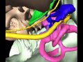 Anatomy of the Facial Nerve in the Temporal Bone with Audio
 Middle ear boundaries simplified with mnemonics
 Middle Ear - Gross Anatomy - Part 4/9
 Anatomy of Eustachian Tube
 Middle Ear - Anatomy | Head and Neck | Agam Webinars
 Mastoid Surgery Animation (Basic to Radical Mastoidectomy)