Visualizing tissues and immune cells in 3D
Animation of a 3D image of intact (non-sectioned) mouse lymph node imaged using the LBS-developed method of Ce3D clearing and staining. Cyan = B220 staining of B cells. Yellow = CD31 staining of vascular endothelial cells. White = LYVE-1 staining of lymphatic endothelial cells. Green = CD8 staining of T cells and CD8+ dendritic cells. Red = CD169 staining of macrophages. Image first shows vascular tree, then adds B cell follicles, then shows in silico computer sectioning of the entire lymph node.
W. LI, M. Gerner, and R. N. Germain, unpublished.
Видео Visualizing tissues and immune cells in 3D канала NIAID
W. LI, M. Gerner, and R. N. Germain, unpublished.
Видео Visualizing tissues and immune cells in 3D канала NIAID
Показать
Комментарии отсутствуют
Информация о видео
Другие видео канала
 Treatment of Patients with DADA2
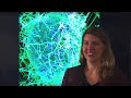 A Q&A with Dr. Erica Ollmann Saphire
 Asthma Advance: How Immune Pathways Contribute to Asthma
 Archived Presentation: NIH HIV/AIDS Network Refinement Protocol Funds Pilot Discussion
 Children Surpass a Year of HIV Remission after Treatment Pause
 Navigating NIAID Welcome Workshop
 NIAID Careers
 Diagnosis and Treatment of the “Undifferentiated Patient,” Part 1
 Difficult-to-Treat CAPS
 The Forgotten Indispensable Man: Joe Kinyoun & The Birth of the NIH
 Biomarker Boot Camp: Interferon Activation
 HIV Advance: Tracking HIV Superinfection Between Partners
 Archived Presentation: Protocol Funds Pilot 1 Implementation Plan
 Better Mad Cow Test
 Boot Camp: Biomarkers in Autoinflammation
 (Audio-Described Version) Alkis Togias, M.D., Discusses the OUtMATCH Trial
 Immunology Advance: Blocking Alarmins to Treat Long-Term Disease
 Addressing the HIV Epidemic in DC
 Discussion: Organizing a Network: Examples from Immunodeficiency Networks
 Movie 15: Sequential migration of neutrophils from the epidermis/dermis into sites of sand fly...