AMYLOIDOSIS: PART 2: Pathogenesis & Classification
The Part 2 of 3 parts tutorial on Amyloidosis.In this part i have described the Pathogenesis and Classification of Amyloidosis.Visit http://ilovepathology.com/ for more topics in Pathology!
****Follow me*****
to view AMyloidosis Part 1 Click here: https://www.youtube.com/watch?v=Icbn9NCYqNc
Twitter : https://twitter.com/VijayPatho
https://twitter.com/ilovepathology2
Facebook: https://www.facebook.com/ilovepathology/
AMYLOIDOSIS 2
PATHOGENESIS: Basic Mechanism
Abnormal folding of proteins/ Misfolded proteins
Mechanism of damage to the tissues
Pressure effect: leading to atrophy
Accumulation in vessel wall : Leading to ischemia and also increased permeability
Direct cytotoxicity: eg, Amyloidogenic light chain accumulation n cardiac cells
Prefibrillar oligomers: are found be more injurious than actual fibrils. In Alzheimer's and ATTR Amyloidosis
Localised amyloidosis
Limited to a single tissue or organ
Can be only microscopic foci or may be evident grossly as nodular masses
The common sites: lung, larynx, skin, bladder, tongue etc.
Microscopic findings: Lymphocytes and plasma cells can be seen in the periphery of amyloid deposits.
Some cases , the amyloid consists of AL protein
Видео AMYLOIDOSIS: PART 2: Pathogenesis & Classification канала ilovepathology
Показать
Комментарии отсутствуют
Информация о видео
Другие видео канала
 AMYLOIDOSIS: PART 3: Morphology, Diagnosis, Special stains, clinical features & Prognosis
 AMYLOIDOSIS: Part 1: Definition, Historical aspects & Properties of Amyloid
 Type I hypersensitivity (IgE-mediated hypersensitivity) - causes, symptoms, pathology
 INFLAMMATION Part 1: General concepts, types , Vascular changes in Acute inflammation
 Hodgkin’s lymphoma | Hodgkin’s Disease | Reed-Sternberg Cell
 PATHOLOGIC CALCIFICATION: Dystrophic & Metastatic Calcification
 What is AL amyloidosis?
 Understanding Amyloidosis - 3D Animation & Overview
 Type II hypersensitivity (cytotoxic hypersensitivity) - causes, symptoms, & pathology
 SLE: Pathogenesis | Dr. Rakesh Nair | Immunology & Rheumatology | Marrow SS Medicine is now live
 Amyloidosis | Image based Revision (PGMEE) | Dr. Devesh Mishra
 NEOPLASIA-1: BASICS: Nomenclature: Benign, Malignant, Mixed tumors, teratoma,Hamartoma, choristoma
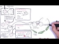 Systemic Lupus Erythematosus (SLE) - signs and symptoms, pathophysiology, investigations, treatment
 NEOPLASIA 3: TUMOR SUPPRESSOR GENES: Retinoblastoma Gene, Knudson's Two Hit Hypothesis.
 APOPTOSIS PART 1: Definition, Causes & Mechanism/Pathways
 NEOPLASIA 4: p53 gene: The Guardian of the genome. functions, regulation and inactivation
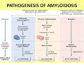 Amyloidosis Part 1
 ALCOHOLIC LIVER DISEASE: Part 1. Pathogenesis
 Cell Injury | General Pathology | Dr. Praveen Kumar Gupta | DBMCI Videos
 HIV & AIDS - signs, symptoms, transmission, causes & pathology