Medial Collateral Ligament Of The Knee - Everything You Need To Know - Dr. Nabil Ebraheim
Dr. Ebraheim's animated educational video describes the medial collateral ligament (MCL) of the knee anatomy and injury.
The medial collateral ligament (MCL) is the most commonly injured ligament of the knee. The MCL is a strong ligament and it fails in valgus. The MCL originates from a sulcus on the distal medial femoral epicondyle. The MCL extends from the medial epicondyle of the femur and inserts into the proximal tibia. The MCL has two parts:
1-Superficial (tibial collateral ligament): deep to the semitendinosus and gracilis tendons. It has a broad insertion 4-6 cm distal to the joint line, deep to the Pes Anserine bursa.
2-Deep medial capsular ligament: it blends with the superficial ligament distally. It is associated with the medial meniscus through the coronary ligament. the posterior fibers of the deep MCL blend with the posteromedial capsule and the posterior oblique ligament.
The POL arises from the medial surface of the femur distal to the adductor tubercle posterior to the origin of the superficial MCL. The POL inserts into the posterior medial corner of the tibia. The POL resists internal tibial rotation in extension.
Superficial (tibial collateral ligament)
•The proximal attachment is on the posterior aspect of the medial femoral condyle approximately 3.2 mm proximal and 4.8 mm posterior to the medial epicondyle of the femur.
•The MPFL originates between the medial epicondyle of the femur and the adductor tubercle (just anterior and distal to the adductor tubercle and superior to the superficial medial collateral ligament).
•The function of the superficial medial collateral ligament, it is the primary restraint to valgus stress at the knee. The length of the superficial medial collateral ligament is about 9.5 cm.
•It has anterior and posterior fibers. The anterior fibers are tighter during 90° of knee flexion. The posterior fibers are tight during knee extension.
•The anterior portion of the medial collateral ligament is the primary stabilizer at 30° of flexion, therefore, to test for injury of the MCL, you will examine the knee in about 30° flexion.
•While testing instability in 30° of flexion is specific for the MCL, testing valgus instability in extension is not specific for MCL injury.
•This is testing the posterior part of the MCL, the posterior oblique ligament, the ACL, the medial posterior capsule and possibly the PCL.
•Clinical exam and findings may be subtle for MCL rupture. Valgus stress test in 30° of flexion may be very helpful. Make sure the leg is free at the edge of the table, so that the test can be done correctly.
An opening of 5 mm or more indicates a complete MCL rupture. Where is the tear?
•Sometimes it is hard to decide and differentiate between an MCL tear and a meniscal tear. Joint line tenderness is specific for a meniscal tear. The McMurray test, the Thessaly test and the Apley compression tests are used to diagnose meniscal tears.
Apley compression test
•If the patient experiences pain with either a click or pop sensation, then the test is positive for a meniscal tear. this test is called Apley compression test because you need to apply compression to the knee joint. In the Apley test, if you rotate the tibia with distractive force which causes the patient pain, then this means that there is a ligamentous injury and not a meniscal tear.
If the MCL is avulsed proximally, there may be tenderness above the joint line and there may be a piece of bone avulsed from the epicondyle. Tear of the MCL in the midsubstance area may be hard to differentiate from a meniscal tear, however if it is avulsed from the tibia distally, then the tenderness is about 6-8 cm from the joint and it acts just like a stener lesion, because the pes anserine tendons mya prevent approximation of the tendons together for healing. This is the one you probably need to fix.
Proximal tear of the MCL from the femur heals better than distal tear which occurs from the tibia.
In the x-rays you can see evidence of the chronic injury.
Pellegrini-Steida Syndrome: Calcification occurring at the origin of the MCL.
MRIs will show the location of the tear.
Treatment
•Usually nonoperative: Ligament usually heals with a scar. Scar matures in 6 weeks to one year. Has about 60% of the strength of a normal medial collateral ligament (MCL)
•Usually treated with hinged knee brace used for 6-8 weeks. Prophylactic hinged knee brace decreases the rate of MCL injuries in contact athletes.
•Combined injury such as ACL and MCL: ACL will be reconstructed after the valgus stability is achieved, except in cases of distal MCL tear, in this case both will be fixed (fix the MCL and reconstruct the ACL).
Follow me on twitter:
https://twitter.com/#!/DrEbraheim_UTMC
Donate to the University of Toledo Foundation Department of Orthopaedic Surgery Endowed Chair Fund:
https://www.utfoundation.org/foundation/home/Give_Online.aspx?sig=29
Видео Medial Collateral Ligament Of The Knee - Everything You Need To Know - Dr. Nabil Ebraheim канала nabil ebraheim
Показать
Комментарии отсутствуют
Информация о видео
Другие видео канала
 Medial Collateral Ligament Injuries - Everything You Need To Know - Dr. Nabil Ebraheim
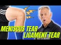 Is Your Knee Pain Coming From a Meniscus Tear or Ligament Strain/Tear? How to Tell.
 MEDIAL & LATERAL COLLATERAL LIGAMENTS ( Anatomy & Biomechanics# applied)
 Tests For Examination Of The Knee - Everything You Need To Know - Dr. Nabil Ebraheim
 LIGAMENTS OF THE KNEE
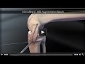 InternalBrace™ MCL Augmentation Repair
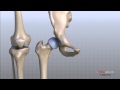 Knee Anatomy Animated Tutorial
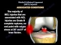 Medial Collateral Ligament injury , MCL Injuries - Everything You Need To Know - Dr. Nabil Ebraheim
 Treatment & Exercises For MCL Strains
 Examination Of L5 Nerve Root - Everything You Need To Know - Dr. Nabil Ebraheim
 Will my ACL, MCL, or PCL (Knee Ligament) Tear Heal?? Surgery??
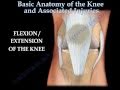 Knee injury ,Injuries - Everything You Need To Know - Dr. Nabil Ebraheim
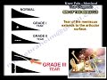 Knee Pain, Meniscal Tear Diagnosis & MRI - Everything You Need To Know - Dr. Nabil Ebraheim
 Top 7 MCL Sprain Treatments - Ask Doctor Jo
 Examination Of L4 Nerve Root - Everything You Need To Know - Dr. Nabil Ebraheim
 A Review Of Acetabular Fractures - Everything You Need To Know - Dr. Nabil Ebraheim
 Valgus Stress Test | Medial Collateral Ligament (MCL) Injury
 Clinical Anatomy - Knee
 Anatomy Of The Knee - Everything You Need To Know - Dr. Nabil Ebraheim
 How to Tell if Knee Pain is Meniscus or Ligament Injury