Cerebrum : Gross anatomy , Relations and External features - Neuroanatomy animations
Join this channel to get access to perks:
https://www.youtube.com/channel/UCG5TBPANNSiKf1Dp-R5Dibg/join
Follow on Instagram:- https://www.instagram.com/drgbhanuprakash
Cerebrum: Gross anatomy, Relations and External features - Neuroanatomy animations
The cerebrum is the largest part of the brain, located superiorly and anteriorly in relation to the brainstem. It consists of two cerebral hemispheres (left and right), separated by the falx cerebri of the dura mater. Embryologically, the cerebrum is derived from the prosencephalon.
Anatomical Position and Structure
--------------------------------------------------------
The cerebrum is located within the bony cranium. It extends from the frontal bone anteriorly to the occipital bone posteriorly. Within the skull, the cerebrum fills the anterior and middle cranial fossae and is located above the tentorium cerebelli anteroposteriorly.
Internal Structure
-----------------------------
The cerebrum is comprised of two different types of tissue – grey matter and white matter:
Grey matter forms the surface of each cerebral hemisphere (known as the cerebral cortex), and is associated with processing and cognition.
White matter forms the bulk of the deeper parts of the brain. It consists of glial cells and myelinated axons that connect the various grey matter areas.
External Structure
------------------------------
Externally, the cerebrum has a highly convoluted appearance, consisting of sulci (grooves or depressions) and gyri (ridges or elevations). It is divided into two anatomically symmetrical hemispheres by the longitudinal fissure – a major sulcus that runs in the median sagittal plane. The falx cerebri (a fold of dura mater) descends vertically to fill this fissure. The two cerebral hemispheres are connected by a white matter structure, called the corpus callosum.
The main sulci are:
------------------------------
Central sulcus – groove separating the frontal and parietal lobes.
Lateral sulcus – groove separating the frontal and parietal lobes from the temporal lobe.
Lunate sulcus – groove located in the occipital cortex.
The main gyri are:
----------------------------
Precentral gyrus – ridge directly anterior to central sulcus, location of primary motor cortex.
Postcentral gyrus – ridge directly posterior to central sulcus, location of primary somatosensory cortex.
Superior temporal gyrus – ridge located inferior to lateral sulcus, responsible for the reception and processing of sound.
Lobes of the Cerebrum
--------------------------------------
The cerebral cortex is classified into four lobes, according to the name of the corresponding cranial bone that approximately overlies each part. Each lobe contains various cortical association areas – where information from different modalities are collated for processing. Together, these areas function to give us a meaningful perceptual interpretation and experience of our surrounding environment.
Frontal Lobe
---------------------
The frontal lobe is located beneath the frontal bone of the calvaria and is the most anterior region of the cerebrum. It is separated from the parietal lobe posteriorly by the central sulcus and from the temporal lobe inferoposteriorly by the lateral sulcus.
The association areas of the frontal lobe are responsible for: higher intellect, personality, mood, social conduct, and language (dominant hemisphere side only).
Parietal Lobe
---------------------
The parietal lobe is found below the parietal bone of the calvaria, between the frontal lobe anteriorly and the occipital lobe posteriorly, from which it is separated by the central sulcus and parieto-occipital sulcus, respectively. It sits superiorly in relation to the temporal lobe, being separated by the lateral sulcus.
Its cortical association areas contribute to the control of: language and calculation on the dominant hemisphere side, and visuospatial functions (e.g. 2-point discrimination) on the non-dominant hemisphere side.
Temporal Lobe
-------------------------
The temporal lobe sits beneath the temporal bone of the calvaria, inferior to the frontal and parietal lobes, from which it is separated by the lateral sulcus.
The cortical association areas of the temporal lobe are accountable for memory and language – this includes hearing as it is the location of the primary auditory cortex.
Occipital Lobe
------------------------
The occipital lobe is the most posterior part of the cerebrum situated below the occipital bone of the calvaria. Its inferior aspect rests upon the tentorium cerebelli, which segregates the cerebrum from the cerebellum. The parieto-occipital sulcus separates the occipital lobe from the parietal and temporal lobes anteriorly.
#cerebrum #cerebrumanatomy #cerebrumanimation #neuroanatomy #cerebrumlecture #usmle #nationalexittest #fmge #mbbs #neuroanatomyvideos #neuroanatomylectures
Видео Cerebrum : Gross anatomy , Relations and External features - Neuroanatomy animations канала Dr.G Bhanu Prakash Animated Medical Videos
Показать
Комментарии отсутствуют
Информация о видео
15 ноября 2020 г. 18:30:12
00:07:26
Другие видео канала
 Basal ganglia Direct and indirect pathways - Neuroanatomy Animations
 Gross Anatomy of the Middle Ear - Boundaries ,Contents and Functions ( Animation )
 Radial nerve Anatomy - Origin, Course, innervation, Saturday night palsy, Wartenberg’s syndrome
 NEUROANATOMY-WHITE MATTER OF CEREBRUM-PART 2-CORPUS CALLOSUM-1-DR ROSE JOSE MD
 Grey matter and white matter (Association fibers, Commissural fibers, Projection fibers ) of CNS
 The Sciatic Nerve Anatomy - Origin, Course, Relations, Branches, Distribution and Clinical anatomy
 Anatomy and Physiology of Larynx , Action of Laryngeal muscles , Dr Bhanu prakash
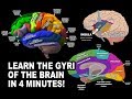 GYRI OF THE BRAIN - LEARN IN 4 MINUTES
 Cerebrum - External Feature
 Circle of Willis : Neuroanatomy Animations
 Internal structures of Cerebral Hemisphere (White/Grey matter) And the Lateral Ventricle - Anatomy
 Neuroanatomy - The Brainstem
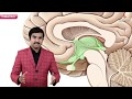 Animated Neuroanatomy : Gross anatomy of Thalamus ( Part 1 ) - Introduction and Relations
 Animated Neuroanatomy : Gross anatomy of Thalamus ( Part 2 ) - Thalamic nuclei
 Introduction: Neuroanatomy Video Lab - Brain Dissections
 Neurology | Gross Anatomy of the Spinal Cord and Spinal Nerves
 NEUROANATOMY-AN INTRODUCTION TO THE WHITE MATTER OF CEREBRUM-PART1-DR ROSE JOSE MD
 Anatomy of the Cerebellum (3D Anatomy Tutorial)
 NEUROANATOMY - CEREBRAL CORTEX PART-1 SULCI AND GYRI - BY DR MITESH DAVE
 Four Lobes of the Brain Mnemonics (Memorable Neurology 1)