GYRI OF THE BRAIN - LEARN IN 4 MINUTES
The brain has four lobes: frontal, parietal, temporal, and occipital. Let’s start examining the gyri of each lobe by first crossing off the ones that are easiest to remember. At the boundary between the frontal and parietal lobes is the central sulcus. The gyrus on the frontal lobe side of this sulcus is the precentral gyrus, while the gyrus on the parietal lobe side is the postcentral gyrus. Next, the frontal and temporal lobes each have a set of three gyri – superior, middle, and inferior. Superior frontal gyrus, middle frontal gyrus, inferior frontal gyrus. Superior temporal gyrus, middle temporal gyrus, inferior temporal gyrus. The occipital lobe is a similar situation, with the superior occipital gyrus, lateral occipital gyrus, and inferior occipital gyrus. Now, let’s get to the gyri that are harder to remember. Here you see the orbital gyri near the eye socket. The inferior frontal gyrus can be split into three sections – the pars orbitalis – closest to your eye socket, or orbit – the pars triangularis – which resembles a triangle – and the pars opercularis… which… who knows why they called it that. Finally, let’s look at the gyri of the parietal lobe. There are the superior and inferior parietal lobules. The inferior parietal lobule is split into the angular gyrus and the supramarginal gyrus.
Now, let’s move onto the medial surface! The gyrus hugging the corpus collosum is the cingulate gyrus. This is the gyrus rectus, or straight gyrus – you’ll see why it’s called that when we examine the brain from the bottom view. This is the superior frontal gyrus. Next, we see the paracentral lobule – so called because within it runs the central sulcus. Then we have the cuneus and precuneus. These two are separated by the parieto-occipital fissure. There is another important fissure called the calcarine fissure. The cuneus lies on its superior bank, and on its lower bank lies the lingual gyrus, so called because it resembles the shape of the tongue. Moving on, we see the parahippocampal gyrus, at the extremity of which lies the uncus. Then there’s the fusiform gyrus, also called the occipitotemporal gyrus, which you use to recognize faces. And then we can see the inferior and middle temporal gyri peaking out.
Let’s look at the brain from the bottom now, and I’ll keep the medial surface visible next to it so you can see where the gyri continue. First there’s the straight gyrus, and now you can see why its called that. At the anterior end of the brain, we see the superior frontal gyrus. Immediately posterior to it are the orbital gyri, bounded laterally by the inferior frontal gyrus. Next, let’s look at the gyri of the temporal lobe. There is the middle temporal gyrus and the inferior temporal gyrus. We also see the fusiform gyrus and the parahippocampal gyrus. Moving onto the occipital lobe, we see the lingual gyrus and the inferior occipital gyrus. We can also see the cingulate gyrus and the cuneus peaking out.
Last view of the brain! Let’s look inside the sylvian fissure, otherwise called the lateral fissure. We see heschl’s gyri, otherwise called the superior transverse temporal gyri here. To better illustrate, here is a coronal cross-section of the brain highlighting the same area. Now, along the top, we see the parietal operculum, the frontal operculum, and the orbital operculum. The inner surface belongs to a structure called the insula (INSULATED INSIDE THE BRAIN), and there are the long gyri of the insula and the short gyri of the insula.
Brain model by:
https://www.turbosquid.com/3d-models/brain-max-free/833681
Видео GYRI OF THE BRAIN - LEARN IN 4 MINUTES канала Neural Academy
Показать
Комментарии отсутствуют
Информация о видео
Другие видео канала
 2-Minute Neuroscience: Lobes and Landmarks of the Brain Surface (Lateral View)
 The Brain
 Blood supply to the brain
 The ventricular system
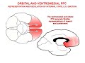 The Neurobiology of Prefrontal Cortex and its Role in Mental Disorders
 Neurology | Cerebral Cortex Anatomy & Function: Overview
 Introduction to Brain Surface Anatomy
 Circulation in Ventricles and Dural Sinuses
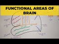 Functional Areas of Brain - 1 | Sulci & Gyri
 How To Fall Asleep In 2 Minutes
 Brain Anatomy 1 - Gross Cortical Anatomy (Lateral Surface)
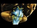 Neuroanatomy - The Brainstem
 The Science of Six Degrees of Separation
 Introduction: Neuroanatomy Video Lab - Brain Dissections
 Cerebral lobes and sulci (basic anatomy)
 Anatomy of the Brain | Dissectible Model
 Temporal Lobe (Cerebral cortex)
 Neurology | Thalamus Anatomy & Function
 Cerebral cortex | Organ Systems | MCAT | Khan Academy
 NEUROANATOMY - CEREBRAL CORTEX PART-1 SULCI AND GYRI - BY DR MITESH DAVE