Osteoid Osteoma - Everything You Need To Know - Dr. Nabil Ebraheim
Dr. Ebraheim’s educational animated video describes the condition of Osteoid Osteoma.
Follow me on twitter:
https://twitter.com/#!/DrEbraheim_UTMC
Osteoid osteoma is a benign, bone forming neoplasm. It has a small nidus of neoplastic tissue surrounded by a heavy zone of reactive mature bone. It is usually located intracortically within the diaphysis of long bones. The proximal femur around the area of the lesser trochanter is a favorite location. The femur and tibia are the most common locations for Osteoid Osteoma to occur in addition to the posterior elements of the spine. It affects the posterior elements of the spine, for example the pedicles or the lamina. Osteoid osteoma is the most common benign tumor of the carpal bones. Osteoid osteoma affects males more than females. 70% of the patients are younger than 20 years old. Osteoid osteoma can look like a stress fracture. If the lesion has large bone reaction, rule out stress fracture. A stress fracture produces more linear radiolucency. With osteoid osteoma, you will have a central lucent nidus area surrounded by a sclerotic area. The nidus is oval or round, and it is well demarcated. The nidus is the lytic lesion. The diameter of the nidus is usually less than 1.5cm. The nidus has a self-limited growth. The osteoid osteoma usually becomes asymptomatic and spontaneously heals. CT scan and MRI will show the lesion as well circumscribed and a cortically based lesion with significant surrounding edema. You will find increased uptake (hot bone scan). Osteoid osteoma is a painful condition that is worse at night and no history of trauma. The painful symptoms are mediated by Prostaglandin E2. There will be increased Cyclooxygenase (COX) activity, which is why the lesion is relieved by aspirin and anti-inflammatory drugs. A differential diagnosis is a Brodie’s Abscess. Osteoid osteoma is located within the cortex. The Brodie’s abscess is located within the medullary canal or in the cancellous bone. The chronic abscess may be surrounded with fibrous tissue and sclerotic bone. It may be difficult to differentiate the Brodie’s abscess from the osteoid osteoma. Other differential diagnoses include osteosarcoma and osteoblastoma. The pathology will show very cellular and vascular stroma with plump, but not atypical osteoblast cells, making a matrix of immature woven bone. The heavy, mature reactive trabeculae encircles the nidus. There will be no inflammatory cells or dead bone to suggest Brodie’s abscess or osteomyelitis. There will be demarcation between the nidus and the bone, and the woven bone will have rimming osteoblasts. Osteoid osteoma is the most common cause of painful scoliosis in young patients. The curvature of the scoliosis is concave towards the site of the lesion. With osteoid osteoma of the thoracic spine, the level of the lesion corresponds to the level of the apex of the resulting scoliosis in the thoracic spine. With osteoid osteoma of the lumbar spine, in the lower lumbar region, the apex of the scoliosis may be above the lesion. Surgical excision of the lesion can help the scoliosis, except if the curvature is large or if excision is delayed in younger patients. The most important step in the treatment will be observation and oral anti-inflammatory medications. This is followed by CT guided radiofrequency ablation if the conservative treatment fails. Radiofrequency ablation (RFA) is usually done in the majority of cases that are painful, except in the spine, because of the proximity to the dura and to the nerve roots, and don’t use RFA in the hand. We don’t use RFA in the digits because of the thermal necrosis of the overlying skin and due to the proximity of the neurovascular structures. The treatment is surgical resection with curettage in the spine and in the digits.
Видео Osteoid Osteoma - Everything You Need To Know - Dr. Nabil Ebraheim канала nabil ebraheim
Показать
Комментарии отсутствуют
Информация о видео
Другие видео канала
 Bone tumors - causes, symptoms, diagnosis, treatment, pathology
 Osteoid Osteoma: Bone Pathology Basics
 Sacral Fractures , Review - Everything You Need To Know - Dr. Nabil Ebraheim
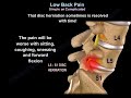 Low Back Pain Simple or Complicated - Everything You Need To Know - Dr. Nabil Ebraheim
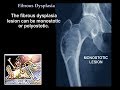 Fibrous Dysplasia - Everything You Need To Know - Dr. Nabil Ebraheim
 Osteoid Osteoma-What we need to know
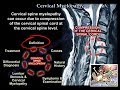 Cervical Myelopathy - Everything You Need To Know - Dr. Nabil Ebraheim
 HUN_2_06_Alkohol okozta mentális és viselkedési zavarok
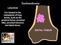 Enchondroma - Everything You Need To Know - Dr. Nabil Ebraheim
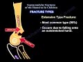 Supracondylar Fractures Of The Humerus In Children
 Bone Tumor : Causes, Symptom, Treatment, Diagnosis, Medical, Surgical & Nursing Management
 Bone Tumors (Benign vs. Malignant)
 Clinical Evaluation of Impingement and Cuff Tears -Everything You Need To Know - Dr. Nabil Ebraheim
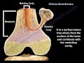 Osteochondroma , solitary and multiple . Everything You Need To Know - Dr. Nabil Ebraheim
 Giant Cell Tumor - Everything You Need To Know - Dr. Nabil Ebraheim
 Acetabulum Fractures Surgical Approaches - Everything You Need To Know - Dr. Nabil Ebraheim
 osteoid osteoma
 Benign Bone Tumors (The Importance & Challenges Of Diagnosis)
 Imaging of Bone Tumors : Key Concepts | Dr. Venkatesh Manchikanti @Shades of Radiology
 Osteonecrosis Of The Hip Stages & Treatment - Everything You Need To Know - Dr. Nabil