The Developmental Tale of the Heart Tube from Cardiogenic Area
🔍 Explore the fascinating journey of the heart tube from the cardiogenic area! 🫀 Join me on a developmental tale that unfolds the secrets of cardiac formation. #HeartTubeDevelopment #CardiologyJourney #EmbryonicHeart #ScienceExploration"
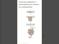
Development of heart tube from cardiogenic area
In the splanchnopleuric mesenchyme on either side of the neural plate, a horseshoe-shaped area develops as the cardiogenic region. This has formed from cardiac myoblasts and blood islands as forerunners of blood cells and vessels.
By day 19, an endocardial tube begins to develop in each side of this region. These two tubes grow and by the third week have converged towards each other to merge, using programmed cell death to form a single tube, the tubular heart
Heart development (also known as cardiogenesis) refers to the prenatal development of the heart. This begins with the formation of two endocardial tubes which merge to form the tubular heart, also called the primitive heart tube. The heart is the first functional organ in vertebrate embryos, and in the human, beats spontaneously by week 4 of development.
The tubular heart quickly differentiates into the truncus arteriosus, bulbus cordis, primitive ventricle, primitive atrium, and the sinus venosus. The truncus arteriosus splits into the ascending aorta and the pulmonary trunk. The bulbus cordis forms part of the ventricles. The sinus venosus connects to the fetal circulation.
The heart tube elongates on the right side, looping and becoming the first visual sign of left-right asymmetry of the body. Septa form within the atria and ventricles to separate the left and right sides of the heart
For anatomy lecture notes and videos : Please visit
1. Difference between : microscopicanatomybd.blogspot.com
https://draft.blogger.com/blog/posts/6434348613242767000
2. Easy humanatomy : easyhumanatomy73.blogspot.com, https://draft.blogger.com/blog/posts/2008939036433143036
For more anatomy creative video please visit
3. easy humanatomy facebook page
https://www.facebook.com/easyhumanatomy
Видео The Developmental Tale of the Heart Tube from Cardiogenic Area канала easy humananatomy
Показать
Комментарии отсутствуют
Информация о видео
Другие видео канала
 Development of endocrine gland
 Landmarks of skull Frankfort line vs Reid's base line
 Histology of vas deferens
 Peritoneal relation is not same considering sex
 General anatomy of nervous system Bangla part 3A
 Bones of head and neck gold information to memorize
 summary of juxtaglomerular apparatus
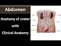 Anatomy of ureter with clinical anatomy
 cartilage histology slides with identifying points #histology #anatomy #biology
 Exploring Norma Lateralis
 Pericardiocentesis I Anatomy of clinical procedure
 Cadaver Exploration of the Heart's Superior Vena Cava
 Difference between mast cells and basophils
 What is the functional importance of elastic artery
 Beyond the Books: Clavicle Viva Explained in our Upper Limb Podcast
 Gray and white rami communicans easy explanation
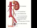 Blood supply of ureter
 Which cell of body do not have a nucleus ?
 Classical hepatic lobule: Architectural Marvel of Liver
 What are the structure passes below the inguinal ligament?
 Parietal Cell of gastric gland