Examination Of The AC Joint - Everything You Need To Know - Dr. Nabil Ebraheim
Dr. Ebraheim’s educational animated video describes the conditions affecting the AC joint.
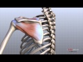
The AC joint is located at the top of the shoulder where the acromion of the scapula and the clavicle join together. The AC joint is a small synovial gliding joint. The AC joint can be affected by arthritis and osteolysis. The oblique orientation of the joint’s articular surfaces may allow the acromion to be driven underneath the clavicle when the AC joint is injured. The condition could be subtle. Injuries of the acromioclavicular joint most commonly occur due to separation of the AC joint. Falling directly onto the shoulder can injure the ligaments that stabilize the AC joint. The AC ligament provides anterior-posterior stability of the AC joint. The posterior and superior AC ligaments are most important for stability. The coracoclavicular ligaments provide superior-inferior stability.
Symptoms: activity related pain with overhead activity and arm adduction.
How to test for injury to the AC joint?
Physical exam: start by palpating the AC joint. Check to see if pain is present with direct palpation of the AC joint. If pressing down onto the AC joint causes pain, this is a sign of an AC joint problem such as distal clavicle osteolysis, arthritis, sprain of the AC ligament or separation.
Osteolysis of the distal clavicle
Localized area of inflammation, hyperemia, microfracture, bone resorption and eventually arthritis of the AC joint.
Provocative tests: when pulling down on the shoulder, if there is separation of the AC joint, the clavicle will rise and a bump will be seen in the area of the joint. Sometimes this is demonstrated by adding weights and comparing both sides. The cross body adduction test can also be done by bringing the shoulder across the body. This squeezes the acromion and clavicle together causing pain directly in the area of the joint if an AC joint separation or arthritis is present.
The acromioclavicular joint is best evaluated using the Zanca view radiograph: direction of the X-ray beam: the beam is directed with cephalad angle of 15 degrees.
Clavicular osteolysis can be assessed using the Zanca view. The acromion will be normal with the abnormality isolated to the distal clavicle. Zanca view is also used for diagnosis of arthritis of the AC joint. It can show osteophytes and joint space narrowing. The patient’s symptoms may not correlate with the x-ray findings. MRI shows increased signal edema in the AC joint.
Become a friend on facebook:
http://www.facebook.com/drebraheim
Follow me on twitter:
https://twitter.com/#!/DrEbraheim_UTMC
Donate to the University of Toledo Foundation Department of Orthopaedic Surgery Endowed Chair Fund:
https://www.utfoundation.org/foundation/home/Give_Online.aspx?sig=29
Background music provided as a free download from YouTube Audio Library.
Song Title: Every Step
Видео Examination Of The AC Joint - Everything You Need To Know - Dr. Nabil Ebraheim канала nabil ebraheim
Показать
Комментарии отсутствуют
Информация о видео
Другие видео канала
 Shoulder Anatomy Animated Tutorial
 The Exam for Shoulder Pain - Stanford Medicine 25
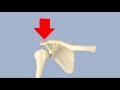 Acromioclavicular Separation
 Shoulder Impingement Syndrome - Everything You Need To Know - Dr. Nabil Ebraheim
 The KEY To Fixing AC Joint Pain
 AC joint separation grade 3 successful rehab protocol
 Shoulder Examination Inspection & Palpation - Everything You Need To Know - Dr. Nabil Ebraheim
 Malunion Of The Clavicle - Everything You Need To Know - Dr. Nabil Ebraheim
 "Weight-lifter's shoulder" pain from an unstable AC joint | Feat. Tim Keeley | No. 27 | Physio REHAB
 Shoulder Pain from Your AC Joint?? 3 Quick Tests You Can Do.
 The Acromioclavicular (AC) Joint | Anatomy and Function
 Fix Your Shoulder Pain (BENCH PRESS!)
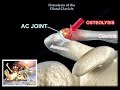 Distal Clavicle Osteolysis - Everything You Need To Know - Dr. Nabil Ebraheim
 What is Causing Your Shoulder Pain? Tests You Can Do Yourself.
 AC Joint Sprain Stretches & Exercises - Ask Doctor Jo
 4 Tests to Differentiate Shoulder Impingement and AC Joint Dysfunction
 AC Joint Pain Exercises for Shoulder Rehab
 Diagnosis of a symptomatic AC Joint | Two Test Clusters
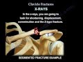 Clavicle Fractures - Everything You Need To Know - Dr. Nabil Ebraheim
 AC Joint Sprain Rehabilitation - Stretches, Exercises and Massage For Faster Recovery