Anatomy Of The Meniscus - Everything You Need To Know - Dr. Nabil Ebraheim
Dr. Ebraheim’s educational animated video describes the Anatomy of the Meniscus.
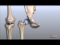
The meniscus is a cushion structure made of cartilage which fits within the knee joint between the tibia and femur. There are two menisci inside the knee joint: medial meniscus and lateral meniscus. The medial meniscus is C-shaped and the lateral meniscus is more circular. The medial meniscus covers 50% of the medial tibial plateau and the lateral meniscus covers 70% of the lateral tibial plateau. With the medial meniscus, the anterior and posterior horns are separated from each other and from the anterior cruciate ligament. The posterior horn of the medial meniscus is the main secondary stabilizer of anterior translation. The anterior and posterior horns of the lateral meniscus are closer to each other and near the insertion of the ACL. The transverse intermeniscal ligament connects the anterior horns of the lateral and medial menisci. The meniscus is made up of type IV collagen. The meniscus provides shock absorption and stability to the knee joint.
The meniscus provides load sharing across the knee by increasing the contact area and decreasing the contact stress. If the meniscus is removed, the patient will develop arthritis of the knee joint. The contact stress increases 2-3 times when the meniscus is removed. The stress increases with the increased loss of meniscal tissue. The meniscus helps to protect the knee joint, allowing the bones to slide freely on each other, however the meniscus limits flexion and extension extremes. The meniscus transmits 50 degrees of force in extension and 90 degrees of force in flexion. Beyond 90 degrees, most of the force is transmitted to the posterior horn of the meniscus (flexion of the knee causes pain).
The blood supply of the meniscus decides the healing potential of the meniscus. The peripheral 1/3 of the meniscus is vascular. It will heal if repaired. The inner 1/3 of the meniscus is not vascular and is nourished by synovial fluid. The middle 1/3 is red/white and it is avascular. The blood supply of the meniscus originates from the medial and lateral geniculate arteries. The meniscus appears triangular in a cross section. Traumatic tear of the meniscus may cause bleeding inside the knee joint. Swelling is usually gradual. It is different from an ACL tear that will have sudden immediate swelling.
The peripheral portion of the meniscus has nerve supply. It is well innervated in the posterior horn (mechanoreceptors). It provides proprioception during joint movement.
Meniscal tears are common involving sports related activities with younger patients. Injury occurs from twisting or jumping activities such as sports like football or skiing. The tear usually is an acute, traumatic injury and it usually requires surgery. In older individuals, tears are degenerative with a gradual onset of pain and the patient may not benefit from surgery. Removal of the meniscus may lead to arthritis of the knee joint.
Tears of the medial meniscus occur 3 times more often than tears of the lateral meniscus. The lateral meniscus is mobile and the medial meniscus is more fixed causing more tears to occur in the medial meniscus compared to the lateral meniscus. The popliteus hiatus makes the lateral meniscus more mobile with less injuries. The lateral meniscus is associated with a discoid meniscus and meniscal cyst. The lateral meniscus is also associated with acute injury to the anterior cruciate ligament.
Tear of the medial meniscus occurs more with degenerative tears with ACL deficient knee. They also are associated with a baker’s cyst.
Anatomical and surgical considerations:
The peroneal nerve is posterior to the biceps femoris tendon and must be protected during repair of the lateral meniscus. The peroneal nerve passes deep to the biceps muscle. The saphenous nerve is posterior to the Sartorius tendon and must be protected during repair of the medial meniscus.
Become a friend on facebook:
http://www.facebook.com/drebraheim
Follow me on twitter:
https://twitter.com/#!/DrEbraheim_UTMC
Donate to the University of Toledo Foundation Department of Orthopaedic Surgery Endowed Chair Fund:
https://www.utfoundation.org/foundation/home/Give_Online.aspx?sig=29
Background music provided as a free download from YouTube Audio Library.
Song Title: Every Step
Видео Anatomy Of The Meniscus - Everything You Need To Know - Dr. Nabil Ebraheim канала nabil ebraheim
Показать
Комментарии отсутствуют
Информация о видео
Другие видео канала
 Knee Anatomy Animated Tutorial
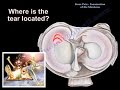 Knee Pain , Meniscus tear - Everything You Need To Know - Dr. Nabil Ebraheim
 Cruciate ligaments of knee ( attachment , function & injury) English , DR. SAMEH GHAZY
 Clinical Anatomy - Knee
 Is Your Knee Pain Coming From a Meniscus Tear or Ligament Strain/Tear? How to Tell.
 Arthroscopic Meniscus Repair of Knee PreOp® Patient Education Feature
 Clinical Anatomy - Knee mensicus and knee joint
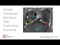 How to Read a Knee MRI for Meniscus Tears
 Systematic Interpretation of Knee MRI: How I do it
 Arthroscopic Meniscus Repair
 Meniscus Tears Part 1 - How can physical therapy help you avoid knee surgery?
 Regenexx Alternative to Knee Meniscus Surgery / Meniscectomy
 Top 7 Exercises after Meniscus Tear (Decrease Pain & Increase Strength)
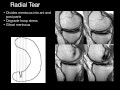 Knee Menisci by Geoffrey Riley, M.D.
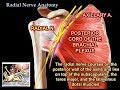 Radial Nerve Anatomy - Everything You Need To Know - Dr. Nabil Ebraheim
 Meniscal tears ... Why are they so common? Is surgery always necessary?
 Top 3 Signs You Have a Meniscus Tear in Your Knee. Tests You Can Do
 ACL, PCL, & Quadriceps - Everything You Need To Know - Dr. Nabil Ebraheim
 Knee Pain , Knee arthritis treatment - Everything You Need To Know - Dr. Nabil Ebraheim, M.D.
 Meniscus Tear Stretches & Exercises - Ask Doctor Jo