Fluoroscopy: Image Intensifier System
Fluoroscopy, or real-time projection X-ray imaging, has been in clinical use since shortly after Roentgen’s discovery of X-rays. Early fluoroscopes consisted simply of an X-ray source and a fluorescent screen, between which the patient was placed. Current fluoroscopy systems fall into two distinct categories: image intensifier and flat-panel detector (FPD). In this lecture, we will focus on the image intensifier based fluoroscopy imaging system.
1. Introduction to Fluoroscopy
2. Image Intensifier: Input Phosphor, Electron Optics, Output Phosphor
3. Video Camera
4. The Optical System
Видео Fluoroscopy: Image Intensifier System канала JP Academia
1. Introduction to Fluoroscopy
2. Image Intensifier: Input Phosphor, Electron Optics, Output Phosphor
3. Video Camera
4. The Optical System
Видео Fluoroscopy: Image Intensifier System канала JP Academia
Показать
Комментарии отсутствуют
Информация о видео
Другие видео канала
 Modes of Radioactive Decay, Decay Schemes, and Series
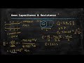 Physics of the Nervous System
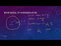 Bohr Atomic Model
 Nuclear Models (Liquid Drop Model & Shell Model) Final
 Introduction to Radioactive Decay
 Current & Resistance
 Nucleus & Chart of Nuclides
 Simple Circuits (Series & Parallel Circuit)
 Atomic Line Spectra and Energy Levels
 Introduction to Vectors
 Overview of Magnetic Resonance Phenomenon & Imaging
 Nuclear Mass & Binding Energy
![[Taglish/Tagalog] Kirchhoff's Rules Example](https://i.ytimg.com/vi/vM1mkr02RHI/default.jpg) [Taglish/Tagalog] Kirchhoff's Rules Example
[Taglish/Tagalog] Kirchhoff's Rules Example Motion in One Dimension (Kinematics & Free Fall)
 Photons & Its Interactions
 Statics (Concepts & Sample Problems)
 Basic Electromagnetism for Magnetic Resonance Imaging
 (Taglish) Introduction to Convolution
 Fundamentals of X ray Production (Characteristic & Bremsstrahlung)
 Introduction to Health Physics