Peripheral Vein Cannulation Using Ultrasound- transverse or longitudinal - which is best
Ultrasound guided vessel cannulation (placement of a needle inside a blood vessel) is an essential skill for modern phlebologists, physicians and surgeons, Veins can be cannulated or accessed using ultrasound in transverse (short) section or longitudinal (long) section. Both methods of ultrasound guided venous cannulation have their advocates. Which imaging method is best? The pros and cons of both methods are discussed and the techniques are illustrated. Ultrasound guided vein cannulation is an important part of the treatment method of varicose veins by VNUS closure, Endovenous laser (evl or elvt), ultrasound guided foam sclerotherapy and Venaseal by Sapheon, These ultrasound guided treatments of veins avoid surgery, general anaesthesia, surgical stripping and hospitalization. Vein treatments performed and guided by ultrasound are associated with rapid recovery, better cosmetic results and lower recurrence rates.
https://www.veincare.academy/
When the vein is imaged in transverse section, it appears as a circle on the screen and the entire circumference of the vein wall as well as the lumen can be visualised. When the vein is imaged in longitudinal section, only the superficial and deep walls of the vein can be seen. When the vein is cannulated in transverse section, the ultrasound transducer (probe) must be moved along the length of the vein to keep the needle tip in view as the needle is advanced. When cannulating the peripheral vein in longitudinal section, the ultrasound transducer is held still over the vein and the entire length of the needle can be seen as it is advanced. Cannuation of the vein in transverse section requires coordinated movement of both hands simultaneously but has the advantage that the lateral walls of the vein can be seen at all times as the needle tip is advanced.
It appears from a discussion with colleagues that the longitudinal imaging method is generally more prevalent among interventional radiologists while vascular surgeons are more likely to use both methods.
Pros of Cannulation in Longitudinal Section: (1) The whole length of the needle is seen as it is advanced; (2) Once the transducer is positioned it is kept still. Cons of Cannulation in Longitudinal Section: (1) Only the superficial and deep walls of the vein are seen (the medial and lateral walls are not in the imaging plane); (2) Tortuous and very small veins can be challenging.
Pros of Cannulation in Transverse Section: (1) Entire circumference of the vein is visualized; (2) Small veins and tortuous veins more easily cannulated; (2) Less likely to "stray" out of lumen by inadvertently puncturing the medial or lateral walls of the vein when advancing the needle. Cons of Cannulation in Transverse Section: (1) Requires coordinated movements of both hands simultaneously (dominant hand advancing needle and non-dominant hand moving transducer to keep the needle tip in view); (2) Higher requirement for good quality imaging equipment.
When assessing deep veins for deep vein thrombosis (DVT) it is important to image in transverse section and when assessing the proximity of important structures to the veins (eg nerves or arteries), transverse imaging is preferred.
https://www.veincare.academy/
Disclaimer: Health professionals should only provide treatments for which they have proper training and knowledge. This video serves only as a source of additional information to healthcare professionals and the public
Видео Peripheral Vein Cannulation Using Ultrasound- transverse or longitudinal - which is best канала The VeinCare Centre
https://www.veincare.academy/
When the vein is imaged in transverse section, it appears as a circle on the screen and the entire circumference of the vein wall as well as the lumen can be visualised. When the vein is imaged in longitudinal section, only the superficial and deep walls of the vein can be seen. When the vein is cannulated in transverse section, the ultrasound transducer (probe) must be moved along the length of the vein to keep the needle tip in view as the needle is advanced. When cannulating the peripheral vein in longitudinal section, the ultrasound transducer is held still over the vein and the entire length of the needle can be seen as it is advanced. Cannuation of the vein in transverse section requires coordinated movement of both hands simultaneously but has the advantage that the lateral walls of the vein can be seen at all times as the needle tip is advanced.
It appears from a discussion with colleagues that the longitudinal imaging method is generally more prevalent among interventional radiologists while vascular surgeons are more likely to use both methods.
Pros of Cannulation in Longitudinal Section: (1) The whole length of the needle is seen as it is advanced; (2) Once the transducer is positioned it is kept still. Cons of Cannulation in Longitudinal Section: (1) Only the superficial and deep walls of the vein are seen (the medial and lateral walls are not in the imaging plane); (2) Tortuous and very small veins can be challenging.
Pros of Cannulation in Transverse Section: (1) Entire circumference of the vein is visualized; (2) Small veins and tortuous veins more easily cannulated; (2) Less likely to "stray" out of lumen by inadvertently puncturing the medial or lateral walls of the vein when advancing the needle. Cons of Cannulation in Transverse Section: (1) Requires coordinated movements of both hands simultaneously (dominant hand advancing needle and non-dominant hand moving transducer to keep the needle tip in view); (2) Higher requirement for good quality imaging equipment.
When assessing deep veins for deep vein thrombosis (DVT) it is important to image in transverse section and when assessing the proximity of important structures to the veins (eg nerves or arteries), transverse imaging is preferred.
https://www.veincare.academy/
Disclaimer: Health professionals should only provide treatments for which they have proper training and knowledge. This video serves only as a source of additional information to healthcare professionals and the public
Видео Peripheral Vein Cannulation Using Ultrasound- transverse or longitudinal - which is best канала The VeinCare Centre
Показать
Комментарии отсутствуют
Информация о видео
Другие видео канала
 How to insert difficult IV or draw blood sample in patients with difficult veins: Best technique
 Ultrasound Guided IV Access
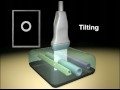 Ultrasound Transducer Manipulation
 Ultrasound-Guided Popliteal Vein Access in Prone Patients
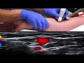 Ultrasound-guided peripheral venous cannulation
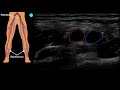 POCUS: Venous Access - Part 1
 Ultrasound Guided Peripheral Access
 Ultrasound guided dynamic needle tip positioning in peripheral vein and artery cannulation
 Peripheral Venous Access Under Ultrasound Guidance - Part 1 - SonoSite, Inc.
 ULTRASOUND GUIDED CANNULATION
 FOCUS ON: Dynamic needle guidance using ultrasound (ICU Point of View minis)
 Traps in Ultrasound Guided Needling
 Sam Hsu MD RDMS Ultrasound-Guided Vascular Access Lecture
 US-Guided Central Line: 10 Steps - Crash course with Dr. Hadzic
 How To: Axillary Vein Cannulation - SonoSite Ultrasound.mp4
 How to Scan the Perforator Veins Episode 4
 Ultrasound Guidance for Central Venous Access - Part 1 - SonoSite, Inc.
 7 steps to cannulate the most difficult veins! Live demonstration
 Ultrasound guided IV placement. 3/2021
 Ultrasound Guided Peripheral IV Placement STEP BY STEP