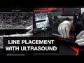
Ultrasound guided dynamic needle tip positioning in peripheral vein and artery cannulation
If you’d like to see the video of an actual patient, visit https://youtu.be/8FnyzJzDYds and view my video: “Ultrasound guided peripheral venous cannulation in an actual patient” .
********* Keyboard Shortcuts(PC) **************
If you feel that the movie is moving too fast, you can control the movie with your computer keyboard as shown below:
Spacebar or K: Toggle Play/Pause.
Comma key:Inch backwards frame by frame during pausing.
Period key: Inch forwards frame by frame during pausing.
Left/Right arrow: Seek backward/forward 5 seconds
J: Seek backward 10 seconds.
L: Seek forward 10 seconds.
********** Contents of this video ***************
Click on the blue type below to jump to a specific stage of the video.
00:00 Intro
00:16 "To-and-fro method"
01:33 Demonstration of the effect of adjusting the incident angle on a simulator
02:32 "To-and-fro method" animation
03:22 "Needle blinking method"
04:38 Determination of the skin entry site
05:00 Demonstration of dynamic ultrasound-guided needle positioning on a simulator
06:51 Prevention of pathogen transmission via ultrasound probe
07:58 Ultrasound-guided needle repositioning
09:38 How to make a home-made simulator
10:59 Take-home Message
*************************************************
As MAGIC guideline suggests, with the aid of ultrasound-guided technique, short catheters and midline catheters should be used more often instead of PICCs and CVCs in the case of difficult vascular access.
The dynamic needle tip positioning technique can be applied to ultrasound-guided cannulation of virtually all peripheral vessels, including hemodialysis arteriovenous fistula, upper arm vein for PICC and midline catheter, and radial artery for monitoring. One exception is calcified arteries (e.g., radial artery of dialysis patients): In such cases, the “needle blinking method” may not work because the ultrasound beam does not penetrate the calcified wall.
A nephrologist in Kyoto, Tadashi Kamata, MD
*************** Relevant YouTube video ***************
How to Safely Cannulate a Difficult Internal Jugular Vein on the First Attempt
https://youtu.be/UfKl2iVbrYs
how to make & practice with an ultrasound phantom for IVs, lines, abscess
https://youtu.be/I-Wqu6Tp8U4
Peripheral Vein Cannulation Using Ultrasound- transverse or longitudinal - which is best
https://youtu.be/RHpzLJHebMU
Ultrasound-Guided Peripheral IV Placement
https://youtu.be/-fduIjQ8EH4
Peripheral Venous Access Under Ultrasound Guidance - Part 1 - SonoSite, Inc.
https://youtu.be/lREUPXCpK8Y
ultrasound guided peripheral IV course by Siegfried Emme
https://youtu.be/d8VFgb9Edfw
Ultrasound-guided arterial line Placement
https://youtu.be/YOxyssqqYNE
PICC LINE INSERTION FULL PROCEDURE
https://youtu.be/9FvUsjje8ic
Видео Ultrasound guided dynamic needle tip positioning in peripheral vein and artery cannulation канала Ultrasound instruction video
Показать
Комментарии отсутствуют
Информация о видео
21 марта 2018 г. 1:24:31
00:11:19
Другие видео канала
 Ultrasound-Guided IV Placement
 Hot Tip-How to Use Ultrasound for Needle Guidance
 16 amazing Water Tricks & Experiments
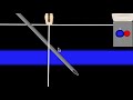 POCUS: Vascular Access - Part 2
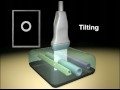 Ultrasound Transducer Manipulation
 Traps in Ultrasound Guided Needling
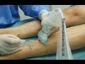 Peripheral Vein Cannulation Using Ultrasound- transverse or longitudinal - which is best
 Central Line Micro-Skills
 Ultrasound Guidance for Central Venous Access - Part 1 - SonoSite, Inc.
 Ultrasound Guided Peripheral Access
 Instructional Series - Sonon Needle Visualization
 FOCUS ON: Dynamic needle guidance using ultrasound (ICU Point of View minis)
 NUTR: Ultrasonic Cutter USW-334
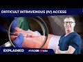 How to insert difficult IV or draw blood sample in patients with difficult veins: Best technique
 IV Insertion: Avoiding Common Mistakes
 ultrasound guided peripheral IV course by Siegfried Emme
 Femoral access: step-by-step and troubleshooting - Mazen Abu-Fadel, MD
 Pneumothorax on Ultrasound - FAST Exam Explained Clearly
 Sam Hsu MD RDMS Ultrasound-Guided Vascular Access Lecture
 Peripheral artery disease: Symptoms