Bilateral Sagittal Splitting Osteotomy - Chang Gung Craniofacial Center
Addressed by Pang-Yun Chou MD
This surgical procedure is performed under general anesthesia and nasotracheal intubation. The face and neck are properly prepped and draped with the whole face exposed, including the forehead. Mandibular osteotomies are performed first. The bilateral sagittal split osteotomy is performed using the modified Hunsuck technique. In 2005, Professor Yu-Ray Chen modified the Hunsuck technique by extending the sagittal osteotomy and making the anterior cut at the first molar (or at least 5 mm proximal to the mental foramen). Increasing sizes of osteotomes are used sequentially to widen the osteotomy site. Subsequently, 2 elevators are used to rock the proximal segment, with the fulcrum against the distal segment. Thus, the 2 segments can be split carefully, thereby reducing the risk of unexpected fracture of the proximal segment. Furthermore, using this technique, the entire mandibular angle can be secured in the proximal segment. When mandibular setback is required, the medial pterygoid and masseter muscles can be detached completely from the medial and lateral aspects of the mandibular angle, respectively. Thus, the mandibular angle included in the proximal segment can be resected or contoured as planned.
The fact that numerous modifications have been made to the original sagittal split ramus osteotomy (SSRO) technique implies that no individual technique can be uniformly satisfactory. In 1957, Obwegeser addressed this technique by involving extensive soft tissue dissection and a horizontal corticotomy on the medial and lateral aspects in the ramus to the posterior border. This technique was found to provide quick bony healing because of the good contact between wide cancellous bone surfaces. However, complications arise with this technique, including excessive swelling and bleeding, damage to the inferior alveolar nerve (IAN), intraoperative trauma to the temporomandibular joint, and unfavorable fractures of the proximal and/or distal segments of the mandible. Dal Pont increased cancellous bone contact using the technique through moving the lateral osteotomy to the distal area of the second molar.
In addition, Hunsuck decreased the extent of osteotomy of the cortical bone on the medial surface of the ramus immediately posterior to the mandibular foramen, which resulted in greater soft-tissue preservation. Epker modified the SSRO to reduce lateral dissection of the masseter and leave major portions of the medial pterygoid and temporalis muscle attached to the proximal segment. He concluded that this modified technique reduced the incidence of excessive swelling and bleeding. In addition, he assumed there would be a lower incidence of infection or avascular necrosis of the proximal segment because of the increased vascularity provided by the attached musculature. Employing concomitant mandibular angle contouring to address a wide lower face resulting from mandibular setback is also possible. However, complications involving mandibular splitting have occurred, such as unexpected fractures, bad splits, facial artery pseudoaneurysms, and facial palsy.
We routinely remove mandibular third molars during BSSO because this may reduce stress experienced by patients and enable greater exposure of impacted third molars using the sagittal split of buccal cortical bone; furthermore, it does not compromise our fixation method, which uses 2-hole plates and screws to cross the anterior osteotomy line.
Therefore, this study concluded that no single SSRO technique fits all cases. Several factors determine the optimal modification of the sagittal split ramus osteotomy. These factors include (1) the position of the mandibular foramen (lingula), (2) the course of the IAN in the mandible, (3) the presence of mandibular third molars, and (4) the planned direction and magnitude of distal segment movement.
As with the rotation of the maxilla discussed in the previous chapter, rotation of the mandible mainly defines the lower facial profile. The lower midline is determined by yaw rotation, and the mandibular plane and gonial prominence are leveled by the mandible’s roll rotation. The pitch rotation of the mandible vertically corrects lip incompetence, adjusts lower facial height, horizontally improves the labiomental fold, and maintains an adequate chin–throat length.
Видео Bilateral Sagittal Splitting Osteotomy - Chang Gung Craniofacial Center канала Pang-Yun Chou
Показать
Комментарии отсутствуют
Информация о видео
Другие видео канала
 Surgical Advancements of the Maxilla and Mandible
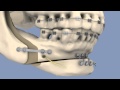 BSSO & Osseous Genioplasty
 CMF - Film patient _ Clivage Sagittal
 BSSO | Easy learning | #stayhome #withme
 ORTHOGNATHIC surgery - All about JAW realignment surgery ©
 CMF - Film patient _ Le Fort 1
 SAGITTAL SPLIT OSTEOTOMY - ORAL SURGERY
 Sagittal Split Osteotomy
 BSSO-Bilateral Sagittal Splitting Osteotomy | 3D Dental Animation
 BSSO | Lower Jaw Advancement Surgery
 Osteotomy Designs | BSSO According To Literature | IPS CaseDesigner® Expert Series
 Anatomy of Temporomandibular joint ( TMJ ) Head and Neck - Gross Anatomy medical animations
 Mandible : Favorite bone of dentists?
 BSSO
BSSO Champy's lines of osteosynthesis
 BSSO - BILATERAL SAGITTAL SPLIT OSTEOTOMY | ORTHOGNATHIC SURGERY | 5 min DENTISTRY easy notes
 LEFORT I OSTEOTOMY || MAXILLARY OSTEOTOMY
 Genioplasty
 Orthognathic Surgery in Kennewick WA: Dr. Cooper | Columbia Basin Oral & Maxillofacial Surgeons
 Johan P. Reyneke — Management (assessment and diagnosis) of the orthognathic patient