Cubital Fossa | Borders & Contents | Anatomy Tutorial
Link to PayPal donation https://paypal.me/studentlamedicina?locale.x=en_US
https://www.instagram.com/anatomy.knowledge/
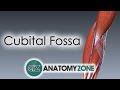
The cubital fossa is a triangular hollow in front of the elbow.
Its floor is formed by two muscles, brachialis in the upper part and supinator in the lower part.
Boundaries of the cubital fossa are formed as follows:
Medially by the lateral margin of pronator teres muscle.
Laterally by the medial margin of brachioradialis muscle.
Superiorly is boundered by an imaginary horizontal line, joining the front of
two epicondyles of the humerus. This imaginary line forms the base of the cubital fossa.
The apex of the fossa is at the meeting point of the lateral and medial boundaries. Here brachioradialis overlaps the pronator teres.
The central anatomical strucure of the cubital fossa is the tendon of biceps brachialis. This tendon is attached to the tuberosity of radius. While crossing form above downward the biceps tendon divides the cubital fossa into a lateral and a medial part.
The medial part contains the following structures:
The brachial artery located imediately medial to the biceps tendon. It terminates in the fossa at the level of neck of radius by dividing into radial and ulnar arteries. The radial artery is superficial and leaves the fossa at the apex. The ulnar artery is deep and leaves the fossa deep to the pronator teres.
The median nerve is placed medial to the brachial artery and leaves the fossa between two heads of pronator teres. The anterior ulnar recurent artery which originates from the superior part of ulnar artery is also present in the medial part of cubital fossa. This artery goes upward ,in the end passing in front of medial epicondyle.
The bicipital aponeurosis originates from the distal insertion of the biceps brachii. While the tendon of the biceps inserts on the radial tuberosity, the aponeurosis reinforces the cubital fossa, and helps to protect the brachial artery and the median nerve running underneath.
The lateral part of cubital fossa most importantly contains the radial nerve. The radial nerve lies in the gap between brachialis and brachioradialis. At the level of lateral epicondyle it divides into two terminal branches: superficial radial nerve and deep radial nerve. The deeps radial nerve dissapears in the substance of supinator muscle while the superficial radial nerve passes downwards under the cover of brachioradialis.
Also present in this region is the radial recurent artery. This artery originates from the radial artery and goes upward in front of the lateral epicondyle.
#cubitalfossa #brachial #mediannerve
Видео Cubital Fossa | Borders & Contents | Anatomy Tutorial канала Anatomy Knowledge
https://www.instagram.com/anatomy.knowledge/
The cubital fossa is a triangular hollow in front of the elbow.
Its floor is formed by two muscles, brachialis in the upper part and supinator in the lower part.
Boundaries of the cubital fossa are formed as follows:
Medially by the lateral margin of pronator teres muscle.
Laterally by the medial margin of brachioradialis muscle.
Superiorly is boundered by an imaginary horizontal line, joining the front of
two epicondyles of the humerus. This imaginary line forms the base of the cubital fossa.
The apex of the fossa is at the meeting point of the lateral and medial boundaries. Here brachioradialis overlaps the pronator teres.
The central anatomical strucure of the cubital fossa is the tendon of biceps brachialis. This tendon is attached to the tuberosity of radius. While crossing form above downward the biceps tendon divides the cubital fossa into a lateral and a medial part.
The medial part contains the following structures:
The brachial artery located imediately medial to the biceps tendon. It terminates in the fossa at the level of neck of radius by dividing into radial and ulnar arteries. The radial artery is superficial and leaves the fossa at the apex. The ulnar artery is deep and leaves the fossa deep to the pronator teres.
The median nerve is placed medial to the brachial artery and leaves the fossa between two heads of pronator teres. The anterior ulnar recurent artery which originates from the superior part of ulnar artery is also present in the medial part of cubital fossa. This artery goes upward ,in the end passing in front of medial epicondyle.
The bicipital aponeurosis originates from the distal insertion of the biceps brachii. While the tendon of the biceps inserts on the radial tuberosity, the aponeurosis reinforces the cubital fossa, and helps to protect the brachial artery and the median nerve running underneath.
The lateral part of cubital fossa most importantly contains the radial nerve. The radial nerve lies in the gap between brachialis and brachioradialis. At the level of lateral epicondyle it divides into two terminal branches: superficial radial nerve and deep radial nerve. The deeps radial nerve dissapears in the substance of supinator muscle while the superficial radial nerve passes downwards under the cover of brachioradialis.
Also present in this region is the radial recurent artery. This artery originates from the radial artery and goes upward in front of the lateral epicondyle.
#cubitalfossa #brachial #mediannerve
Видео Cubital Fossa | Borders & Contents | Anatomy Tutorial канала Anatomy Knowledge
Показать
Комментарии отсутствуют
Информация о видео
Другие видео канала
 Cubital Fossa | 3D Anatomy Tutorial
 Neurology | Brachial Plexus
 Cubital Fossa - Everything You Need To Know - Dr. Nabil Ebraheim
 Thigh Muscles - Medial Compartment of Thigh | Anatomy Tutorial
 Radial and Ulnar arteries - Course & Branches | Anatomy Tutorial
 CUBITAL FOSSA || Boundaries || Contents || Clinical Importance | Easy Explaination
 Elbow Joint: Bones, Muscles & Movement - Human Anatomy | Kenhub
 Superficial Veins of Upper Limb - Basilic & Cephalic veins | Anatomy Tutorial
 Cubital fossa anatomy - Boundaries, contents, and clinical anatomy
 Popliteal Fossa
 Radial Nerve - part #1 | Anatomy Tutorial
 Brachial Artery and its branches - Anatomy Tutorial
 Cubital Fossa || Usmle Cadaveric Anatomy - Boundaries and Contents
 AXILLARY ARTERY ANATOMY ANIMATED LECTURE
 Great Saphenous Vein & Small Saphenous Vein - Venous drainage of lower limb
 BRACHIAL PLEXUS - Anatomy Tutorial
 Muscles of the Upper Limb
 3D Tour of the Axilla
 Shoulder joint Anatomy - Ligaments, Movements, Blood supply , Nerve supply and Clinical anatomy
 Anterior Forearm Muscles | Superficial Layer