Cubital fossa anatomy - Boundaries, contents, and clinical anatomy
►𝐉𝐨𝐢𝐧 𝐓𝐡𝐢𝐬 𝐂𝐡𝐚𝐧𝐧𝐞𝐥 𝐓𝐨 𝐆𝐞𝐭 𝐀𝐜𝐜𝐞𝐬𝐬 𝐓𝐨 𝐏𝐞𝐫𝐤𝐬 :- https://bit.ly/2RQHvTN
►𝐃𝐨𝐰𝐧𝐥𝐨𝐚𝐝 𝐭𝐡𝐞 𝐌𝐞𝐝𝐯𝐢𝐳𝐳 𝐚𝐩𝐩 𝐮𝐬𝐢𝐧𝐠 𝐭𝐡𝐞 𝐛𝐞𝐥𝐨𝐰 𝐥𝐢𝐧𝐤 👇👇👇👇 𝐃𝐨𝐰𝐧𝐥𝐨𝐚𝐝 👇👇👇👇
►𝐀𝐧𝐝𝐫𝐨𝐢𝐝 :- https://bit.ly/3ansFKq
📌𝐅𝐨𝐥𝐥𝐨𝐰 𝐨𝐧 𝐈𝐧𝐬𝐭𝐚𝐠𝐫𝐚𝐦 :-
https://www.instagram.com/drgbhanuprakash
Cubital fossa anatomy - Boundaries, contents, and clinical anatomy
The cubital fossa is a triangular-shaped area or depression situated in relation to the ventral surface of the forearm and contains important neurovascular structures. It has a superior, medial and lateral border, as well as an apex that is directed inferiorly. The cubital fossa also has a floor and roof, and it is traversed by structures that make up its contents.
Boundaries
------------------
Base: imaginary line joining the epicondyles of the humerus
Medial border: pronator teres muscle
Lateral border: brachioradialis muscle
Apex: pronator teres and brachioradialis muscles
Roof: skin, fascia of forearm, bicipital aponeurosis
Floor: brachialis muscle, supinator muscle
Contents
---------------
The median nerve, Brachial artery, Tendon of biceps brachii, Radial nerve
Mnemonic: My Blood Turns Red
Clinical points Venipuncture, blood pressure measurements
#cubitalfossa #anatomyofcubitalfossa #contentsofcubitalfossa #boundariesofcubitalfossa #anatomy #usmle #fmge #neetpg
Видео Cubital fossa anatomy - Boundaries, contents, and clinical anatomy канала Dr.G Bhanu Prakash Animated Medical Videos
Показать
Комментарии отсутствуют
Информация о видео
21 сентября 2018 г. 19:44:21
00:03:46
Другие видео канала
 Cubital Fossa - Everything You Need To Know - Dr. Nabil Ebraheim
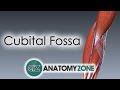 Cubital Fossa | 3D Anatomy Tutorial
 Anatomy of Brachial Artery - Origin , Course , Branches and Relations - USMLE , FMGE and Neet PG
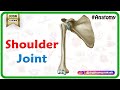 Shoulder joint Anatomy - Ligaments, Movements, Blood supply , Nerve supply and Clinical anatomy
 Clavipectoral fascia gross anatomy - Extension , attachments , structures piercing medical animation
 Radial nerve Anatomy USMLE Origin, Course, innervation, Saturday night palsy, Wartenberg’s syndrome
 Rotator cuff muscles Anatomy - HD Animation
 Shoulder joint: Movements, bones and muscles - Human Anatomy | Kenhub
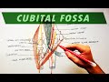 Cubital Fossa | Borders & Contents | Anatomy Tutorial
 CUBITAL FOSSA || Boundaries || Contents || Clinical Importance | Easy Explaination
 Ulnar nerve Anatomy: Origin, Course, Branches, Cubital Tunnel Syndrome || #Usmle #neetpg
 Anatomy Of The Popliteal Fossa - Everything You Need To Know - Dr. Nabil Ebraheim
 Upper Limb Arteries - Arm and Forearm - 3D Anatomy Tutorial
 Brachial plexus ( Roots , Trunks , Divisions , Cords and Branches ) - Origin, Course , Relations
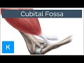 Cubital Fossa - Location & Borders - Human Anatomy | Kenhub
 The Carpal Tunnel easiest way to learn! - Anatomy
 Pleura (anatomy)
 Elbow Joint: Bones, Muscles & Movement - Human Anatomy | Kenhub
 AXILLARY ARTERY ANATOMY ANIMATED LECTURE
 Great saphenous vein - Animated Gross anatomy of lower limb ( Courtery : Dr vishram singh )