How to Read Knee MRI of MCL Injury | Complex Knee Surgeon | Knee Pain Diagnosis | Twin Cities, MN
http://drrobertlaprademd.com/
Complex knee surgeon, Robert LaPrade MD, PhD. breaks down how to read knee MRI of MCL injury. This patient had a skiing injury on the medial side of the knee.
To begin, the coronal image is assessed. In this type of image the normal structures will be dark and the torn structures will be light. A lot of the white indicates a majority of the swelling on the medial side of the knee.
We then move to the sagittal images. As you move more midline you can begin to see more swelling form the injury. We can see the ACL, which in this case is still intact. We also see a complete PCL tear between the tibial attachment site and the femoral attachment site. You can see the that menisci are still intact.
The last image that we will use is the axial image. One of the first things we see where the MCL tibial attachment would be and all of the edema and swelling surrounding that area. We see a hint of the MCL attachment site still attached on the femur, but it has otherwise been completely disrupted.
#mclmri #kneesurgery #kneediagnosis
To learn more about how to read knee MRI of MCL injuries, please visit: https://drrobertlaprademd.com/mcl-injuries-medial-ligament-injury-medial-knee-reconstruction-twin-cities-minnesota/
To learn more, go to:
► Website: https://drrobertlaprademd.com/
► Contact us: https://drrobertlaprademd.com/contact-us/
► Facebook: https://www.facebook.com/kneespecialist
► Twitter: https://twitter.com/thekneedoc
► Instagram: https://www.instagram.com/thekneedoc/
► LinkedIn: https://www.linkedin.com/in/drrobertlaprade/
Dr. LaPrade, MD, PhD has specialized skills and expertise in diagnosing and treating complicated knee injuries. He has treated athletes at all levels, including Olympic, professional and intercollegiate athletes, and has returned numerous athletes back to full participation after surgeries. Recognized globally for his outstanding and efficient surgical skills and dedication to sports medicine, he has received many research awards, including the OREF Clinic Research Award considered by many a Nobel Prize in orthopedics. Dr. LaPrade is one of the most published investigators in his field, and many of the surgeries that he has developed are now performed worldwide and recognized as the “gold standard” for the treatment of complex knee injuries.
Видео How to Read Knee MRI of MCL Injury | Complex Knee Surgeon | Knee Pain Diagnosis | Twin Cities, MN канала Robert LaPrade
Показать
Комментарии отсутствуют
Информация о видео
Другие видео канала
 Knee MRI scan - Medial Collateral Ligament Anatomy (MCL and medial meniscus tear)
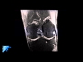 How to Read Knee MRI of Normal Knee | Anatomy of the Knee | Complex Knee Surgeon | Minneapolis , MN
 How to Read an MRI of a PCL Tear | PCL Injury Treatment Options | Sports Medicine | Twin Cities, MN
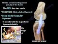 Medial Collateral Ligament Of The Knee - Everything You Need To Know - Dr. Nabil Ebraheim
 How to Tell if Knee Pain is Meniscus or Ligament Injury
 How to Read Knee MRI of LCL Tear | Complex Knee Surgeon | Posterolateral Corner Injury Minnesota, MN
 How to Read Knee MRI of Radial Meniscus Tear | Sports Medicine Knee Specialist | Twin Cities, MN
 How to Read an MRI of the Brain | First Look MRI
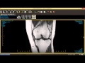 How to Read Knee MRI
 MCL Sprain Rehab Exercise Progressions | Early, Mid-, & Late Stage
 Treatment & Exercises for MCL Strains
 Systematic Interpretation of Knee MRI: How I do it
 Rotator Cuff tear Imaging - Everything You Need To Know - Dr. Nabil Ebraheim
 How to Read Knee MRI of ACL Tear | Anterior Cruciate Ligament Pain | Knee Surgery | Minneapolis, MN
 MCL Injury Rehab excercises
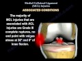 Medial Collateral Ligament injury , MCL Injuries - Everything You Need To Know - Dr. Nabil Ebraheim
 Systematic Interpretation of Knee MRI: Supplemental Cases
 Knee Pain, Meniscal Tear Diagnosis & MRI - Everything You Need To Know - Dr. Nabil Ebraheim
 How to Read Knee MRI of Osteochondritis Dissecans Lesion | OCD Lesions | Knee Pain | Minneapolis, MN
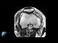 PLC Injury Knee MRI | Posterolateral Corner Knee Injury | Knee Pain Symptoms | Minneapolis, MN