How to Read Knee MRI of Normal Knee | Anatomy of the Knee | Complex Knee Surgeon | Minneapolis , MN
http://drrobertlaprademd.com/
Complex knee specialist Dr. Robert LaPrade discusses how to read knee MRI of normal knee. Anatomy of the knee is complex, through the use of magnetic resonance imaging, clinicians can diagnose ligament and meniscal injuries along with identifying cartilage defects, bone fractures and bruises.
To begin, we use a coronal scan of a left knee. A coronal scan goes through the knee, front to back, with the dimensions being medial to lateral. To begin a coronal scan the patella is at the center. As the image moves deeper you can begin to see the quadriceps tendon and the patellar tendon below. We then move to the tibiofemoral joint. The medial and lateral meniscus come into view and the IT band on the side.
As the image moves deeper you can begin to see the medial collateral ligament along the tibia and the anterior curciate ligament. On the lateral side of the knee the fibular collateral ligament. The ACL becomes more clear as you more deeper into the knee. The medial and lateral meniscal root attachments begin to appear. As we move more posterior aspect of the knee you can see the biceps femoris. Clinically, the coronal view is used to identify any medial or lateral meniscus injuries.
The next image used is the sagittal views. The sagittal views look at the knee from front to back. You can clearly see the posterior and anterior horn of the lateral meniscus. The gray area around the bone is the articular cartilage. As we get more central in the joint, we can begin to see the patellar tendon. The ACL and PCL also come into view. As we move to the far medial aspect we will start to see the hamstring tendons.
The last view is the axial view, which is like cutting through a log. As the image moves deeper into the knee you can see the outline of the menisci. We look for any ghost signs that may appear for radial tears. You can see the shiny white fibers of the posterior horn of the medial meniscus.
To learn more about how to read knee MRI of normal knee and knee injuries, please visit: http://drrobertlaprademd.com/injuries.
#kneemri #kneeanatomy #kneediagnosis
To learn more, go to:
► Website: https://drrobertlaprademd.com/
► Contact us: https://drrobertlaprademd.com/contact-us/
► Facebook: https://www.facebook.com/kneespecialist
► Twitter: https://twitter.com/thekneedoc
► Instagram: https://www.instagram.com/thekneedoc/
► LinkedIn: https://www.linkedin.com/in/drrobertlaprade/
Dr. LaPrade, MD, PhD has specialized skills and expertise in diagnosing and treating complicated knee injuries. He has treated athletes at all levels, including Olympic, professional and intercollegiate athletes, and has returned numerous athletes back to full participation after surgeries. Recognized globally for his outstanding and efficient surgical skills and dedication to sports medicine, he has received many research awards, including the OREF Clinic Research Award considered by many a Nobel Prize in orthopedics. Dr. LaPrade is one of the most published investigators in his field, and many of the surgeries that he has developed are now performed worldwide and recognized as the “gold standard” for the treatment of complex knee injuries.
Видео How to Read Knee MRI of Normal Knee | Anatomy of the Knee | Complex Knee Surgeon | Minneapolis , MN канала Robert LaPrade
Показать
Комментарии отсутствуют
Информация о видео
Другие видео канала
 Medial Meniscus Assessment | Orthopedic Knee Doctor | Meniscus Treatment Options | Twin Cities, MN
 Figure 4 Test Knee | Meniscal Knee Tears | Best Knee Surgeon In The World | Minneapolis St Paul, MN
 Fresh Osteoarticular Allografts | Treatment of Osteochondral Defects Knee | Minneapolis, MN
 Lateral Patellar Translation | Clinical Knee Exam | Patellofemoral Pain Syndrome | Twin Cities, MN
 Knee Surgery Patient Story | Complex Knee Injury | Sports Medicine and Injury Care | Twin Cities, MN
 Knee Extension Exercises | Knee Rehabilitation Program | Knee Injury Treatment | Minneapolis, MN
 Pivot Shift Test Knee | Sports Medicine Knee Doctor | Orthopedic Surgeon | Minneapolis St Paul, MN
 Anterior Drawer Test | Clinical Knee Exam | Knee Injury Diagnosis | Minneapolis Saint Paul, MN
 Swollen Thigh After Injury | Varus Stress Test Knee | Orthopedic Knee Surgeon | Minneapolis, MN
 Knee Valgus Stress Test | Clinical Knee Examination | Knee Extension of Degrees | Minneapolis MN
 Quadriceps Exercise After Knee Injury | Bad Knee Pain Relief | Knee Specialist | Minneapolis, MN
 Assessment of Knee Range of Motion | Knee Pain Relief Exercises | Knee Injury | Minneapolis, MN
 Patellar Translation Measurement | Knee Exam Test | MRI of Knee Injury | Knee Pain | Minneapolis, MN
 Varus Stress Test Knee Exam | Sports Medicine | Orthopedic Knee Surgeon | Minneapolis St Paul, MN
 Anterolateral Ligament | All Ligaments In The Knee | ACL Tear After Reconstruction | Minneapolis, MN
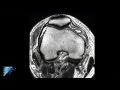 PLC Injury Knee MRI | Posterolateral Corner Knee Injury | Knee Pain Symptoms | Minneapolis, MN
 How to Read Knee MRI of Osteochondritis Dissecans Lesion | OCD Lesions | Knee Pain | Minneapolis, MN
 What is a Meniscus Root Tear? | Why Repair a Meniscus Root Tear? | Knee Surgeon | Minneapolis, MN
 Knee Dissection Video | Knee Surgery and Related Research | Knee Doctor | Minneapolis St Paul, MN
 Knee Osteoarthritis Treatment and Symptoms | ACL Reconstruction Surgery Recovery| Minneapolis, MN
 How to Read Knee MRI of LCL Tear | Complex Knee Surgeon | Posterolateral Corner Injury Minnesota, MN