Anatomy of the Pronator Teres Muscle - Everything You Need To Know - Dr. Nabil Ebraheim
Dr. Ebraheim’s educational animated video describes the anatomy of the pronator teres muscle.
Follow me on twitter:
https://twitter.com/#!/DrEbraheim_UTMC
Anatomy of the Pronator Teres Muscle
The pronator teres is a muscle located in the forearm. The pronator teres muscle has two heads. The superficial head, which is the humeral head, arises from the medial epicondyle of the humerus and a deep head, which is the ulnar head, which arises from the medial border of the coronoid process of the ulna. The muscle is inserted into the middle of the lateral surface of the shaft of the radius. The function of the pronator teres is to pronate the forearm (along with the pronator quadratus) so that the palm turns posterior or down. It functions to pronate the forearm. The pronator teres also assists in flexion of the forearm at the elbow. When fractures occur below the insertion of the pronator teres muscle, the proximal fragment is usually pulled into pronation. The forearm has a normal range of 160-180 degrees. Sometimes a decrease in rotation motion by 50 degrees may not be noticed; however, every attempt should be made to reduce the forearm fracture well, especially fracture of the proximal radius (prone to malrotation). The bicipital tuberosity of the radius is used as a guide to the rotation of the proximal radius. Compare the position of the thumb to the position of the radial tuberosity. The pronator teres muscle is innervated by the median nerve. In the condition of high radial nerve palsy is helped by transfer of the pronator teres muscle. Extension of the wrist, which is impaired by high radial nerve palsy (wrist drop), is improved by transfer of the pronator teres to the extensor carpi radialis brevis muscle. The median nerve passes between the two heads of the pronator teres. The ulnar head (deep hard) separates the median nerve from the ulnar artery. The median nerve runs above the deep head while the ulnar artery runs below the deep head. It seems like when a nerve enters the forearm, it goes between two heads of a muscle. The posterior interosseous nerve enters the extensor compartment of the forearm between the two heads of the supinator muscle. The ulnar nerve enters the forearm by passing through the two heads of the flexor carpi ulnaris muscle. The median nerve enters the forearm by passing through the two heads of the pronator teres muscle. The condition of pronator teres syndrome can occur due to entrapment of the median nerve between the two heads of the pronator teres muscle. Other causes may cause pronator teres syndrome, however, entrapment of the median nerve between the two heads of the pronator teres is an important cause.
Видео Anatomy of the Pronator Teres Muscle - Everything You Need To Know - Dr. Nabil Ebraheim канала nabil ebraheim
Показать
Комментарии отсутствуют
Информация о видео
Другие видео канала
 Pronator Teres Syndrome - Everything You Need To Know - Dr. Nabil Ebraheim
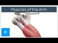 Muscles of the arm - Origin, Insertion & Innervation - Human Anatomy | Kenhub
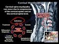 Cervical Myelopathy - Everything You Need To Know - Dr. Nabil Ebraheim
 Anatomy Of The Pronator Quadratus - Everything You Need To Know - Dr. Nabil Ebraheim
 Pronator Teres Stretch: Elbow in Extension
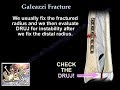 Galeazzi Fracture - Everything You Need To Know - Dr. Nabil Ebraheim
 Acromioclavicular Joint Injury, AC separation - Everything You Need To Know - Dr. Nabil Ebraheim
 Rotator Cuff tear Imaging - Everything You Need To Know - Dr. Nabil Ebraheim
 Pronator teres muscle in less than 1 minute - Kenhub #shorts
 Osteomyelitis Bone Infection - Everything You Need To Know - Dr. Nabil Ebraheim
 Pronator Teres | Muscle Anatomy | Doctor Speaks
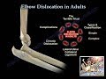 Elbow Dislocation In Adults - Everything You Need To Know - Dr. Nabil Ebraheim
 Pronator Quadratus Muscle Function & Location - Human Anatomy | Kenhub
 Guyan's Canal Syndrome - Everything You Need To Know - Dr. Nabil Ebraheim
 Large shoulder muscles
 Thumb Muscle Anatomy (With Movements!)
![Branches of the Ulnar & Median Nerves [in Hand]](https://i.ytimg.com/vi/ElRwFzT_I58/default.jpg) Branches of the Ulnar & Median Nerves [in Hand]
Branches of the Ulnar & Median Nerves [in Hand] Flexor Digitorum Superficialis - Everything You Need To Know - Dr. Nabil Ebraheim
 Fracture Of The Radial Head Essex Lopresti - Everything You Need To Know - Dr. Nabil Ebraheim
 Plantaris Tendon Rupture - Everything You Need To Know - Dr. Nabil Ebraheim