Osteomyelitis Bone Infection - Everything You Need To Know - Dr. Nabil Ebraheim
Dr. Ebraheim’s educational animated video describes osteomyelitis bone infection.
Follow me on twitter:
https://twitter.com/#!/DrEbraheim_UTMC
Osteomyelitis Bone Infection
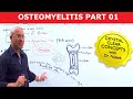
Osteomyelitis is an infection of the bone. It may be an incurable disease. Usually bacteria causes infection in the bone. Staph aureus is the most common organism in adults. Leukocytes are attracted to the area and secrete enzymes in attempt to kill the bacteria. Blood flow to the area is decreased and a devitalized, necrotic bone is formed called a sequestrum. A sequestrum is infected, dead bone resulting from osteomyelitis. Haversian canals surround blood vessels and nerve cells throughout the bone. The sequestrum has no connection to normal bone through the Haversian system (osteon). Because of the fact that the sequestrum is avascular or a dead piece of bone, antibiotics cannot reach the sequestrum or the bacteria. In fact, the bacteria enters the bone cells and hides inside them. Antibiotics alone may not help due to difficulty in penetrating the necrotic area. The involucrum is new bone formation around the sequestrum. The body is trying to seal off the infection by forming new bone. The sequestrum will drain through the sinus. Biopsy of the sinus is not representative of the infection. Multiple deep samples, preferably bone biopsy and cultures, are needed. Biopsy of the sinus is important in long standing cases of osteomyelitis to rule out the formation of squamous cell carcinoma. Associated medical conditions include dialysis, malnutrition, diabetes, IV drug use, and immunosuppression. Patients with sickle cell anemia may have osteomyelitis caused by Salmonella; however, Staph aureus is the most common cause. Patients with IV drug use that have acromioclavicular or sternoclavicular joint infections may occur due to Pseudomonas. The patient may also get pseudomonas from a puncture wound through the shoes. Immunosuppressed patients and patients on parental nutrition may get fungal osteomyelitis (very rare). In children, Eosinophilic granuloma, Ewing’s sarcoma, and acute osteomyelitis may resemble each other. The patient may have pain, fever, or tenderness of the area, and the patient may also have an increased sedimentation rate and leukocytosis. Osteomyelitis can also be confused with a healing fracture or with a benign or malignant tumor. Sometimes a biopsy is necessary for the diagnosis. Only 50% of chronic musculoskeletal infections will have elevated inflammatory markers. Osteomyelitis can be acute, chronic, or subacute. Acute osteomyelitis is usually within 2 weeks. Chronic osteomyelitis is after several months. Subacute is from 4 weeks to several months. There are three types of patients and four types of bone infections in osteomyelitis. The three types of patients are healthy, compromised, and severe systemic compromise. Compromised patients can be locally compromised when they have sinus tract, free flap, and decreased blood supply or systemically compromised when they have comorbidities. In patients with severe systemic compromise, the host in whom treatment will lead to greater morbidity than the infection itself. The four types of bone infections are medullary, superficial, localized defect with stable bone, and diffuse infection with involvement of bone stability. Treatment of osteomyelitis is usually a combination of surgical debridement of the necrotic, nonviable tissue plus administration of culture specific antibiotics. Fist, open the involucrum, next remove the sequestrum (dead bone), then saucerize the bone. Make sure a pathological fracture is not created. Stabilize the bone if needed (external fixator is usually preferred). Fill the cavity with bone chips, cement, or a muscle flap if needed. Intravenous antibiotics are usually given for a period of 6 weeks (usually organism specific). Recurrence of infection is high and occurs in about 30% of cases. MRSA osteomyelitis occurs with a body temperature more than 38 degrees, a WBC count more than 12,000, a hematocrit less than 34%, and a C-reactive protein more than 13. These four independent predictors differentiate between MRSA and MSSA osteomyelitis with 92% chance of having MRSA if all the four are present. If you have MRSA, you give vancomycin or clindamycin. In MRSA, you will have a higher incidence of DVT than other causes of osteomyelitis. Older children, 8 years old or more, with MRSA osteomyelitis and CRP more than 6 have a 40% incidence of DVT on presentation. The presence of Panton-valentine leucocidin gene encoded in strains of MRSA bacteria may explain the deep venous thrombosis (DVT). Careful workup and staging of the bone and the host utilizing the Cierny-Mader classification is important to develop a successful treatment plan.
Видео Osteomyelitis Bone Infection - Everything You Need To Know - Dr. Nabil Ebraheim канала nabil ebraheim
Follow me on twitter:
https://twitter.com/#!/DrEbraheim_UTMC
Osteomyelitis Bone Infection
Osteomyelitis is an infection of the bone. It may be an incurable disease. Usually bacteria causes infection in the bone. Staph aureus is the most common organism in adults. Leukocytes are attracted to the area and secrete enzymes in attempt to kill the bacteria. Blood flow to the area is decreased and a devitalized, necrotic bone is formed called a sequestrum. A sequestrum is infected, dead bone resulting from osteomyelitis. Haversian canals surround blood vessels and nerve cells throughout the bone. The sequestrum has no connection to normal bone through the Haversian system (osteon). Because of the fact that the sequestrum is avascular or a dead piece of bone, antibiotics cannot reach the sequestrum or the bacteria. In fact, the bacteria enters the bone cells and hides inside them. Antibiotics alone may not help due to difficulty in penetrating the necrotic area. The involucrum is new bone formation around the sequestrum. The body is trying to seal off the infection by forming new bone. The sequestrum will drain through the sinus. Biopsy of the sinus is not representative of the infection. Multiple deep samples, preferably bone biopsy and cultures, are needed. Biopsy of the sinus is important in long standing cases of osteomyelitis to rule out the formation of squamous cell carcinoma. Associated medical conditions include dialysis, malnutrition, diabetes, IV drug use, and immunosuppression. Patients with sickle cell anemia may have osteomyelitis caused by Salmonella; however, Staph aureus is the most common cause. Patients with IV drug use that have acromioclavicular or sternoclavicular joint infections may occur due to Pseudomonas. The patient may also get pseudomonas from a puncture wound through the shoes. Immunosuppressed patients and patients on parental nutrition may get fungal osteomyelitis (very rare). In children, Eosinophilic granuloma, Ewing’s sarcoma, and acute osteomyelitis may resemble each other. The patient may have pain, fever, or tenderness of the area, and the patient may also have an increased sedimentation rate and leukocytosis. Osteomyelitis can also be confused with a healing fracture or with a benign or malignant tumor. Sometimes a biopsy is necessary for the diagnosis. Only 50% of chronic musculoskeletal infections will have elevated inflammatory markers. Osteomyelitis can be acute, chronic, or subacute. Acute osteomyelitis is usually within 2 weeks. Chronic osteomyelitis is after several months. Subacute is from 4 weeks to several months. There are three types of patients and four types of bone infections in osteomyelitis. The three types of patients are healthy, compromised, and severe systemic compromise. Compromised patients can be locally compromised when they have sinus tract, free flap, and decreased blood supply or systemically compromised when they have comorbidities. In patients with severe systemic compromise, the host in whom treatment will lead to greater morbidity than the infection itself. The four types of bone infections are medullary, superficial, localized defect with stable bone, and diffuse infection with involvement of bone stability. Treatment of osteomyelitis is usually a combination of surgical debridement of the necrotic, nonviable tissue plus administration of culture specific antibiotics. Fist, open the involucrum, next remove the sequestrum (dead bone), then saucerize the bone. Make sure a pathological fracture is not created. Stabilize the bone if needed (external fixator is usually preferred). Fill the cavity with bone chips, cement, or a muscle flap if needed. Intravenous antibiotics are usually given for a period of 6 weeks (usually organism specific). Recurrence of infection is high and occurs in about 30% of cases. MRSA osteomyelitis occurs with a body temperature more than 38 degrees, a WBC count more than 12,000, a hematocrit less than 34%, and a C-reactive protein more than 13. These four independent predictors differentiate between MRSA and MSSA osteomyelitis with 92% chance of having MRSA if all the four are present. If you have MRSA, you give vancomycin or clindamycin. In MRSA, you will have a higher incidence of DVT than other causes of osteomyelitis. Older children, 8 years old or more, with MRSA osteomyelitis and CRP more than 6 have a 40% incidence of DVT on presentation. The presence of Panton-valentine leucocidin gene encoded in strains of MRSA bacteria may explain the deep venous thrombosis (DVT). Careful workup and staging of the bone and the host utilizing the Cierny-Mader classification is important to develop a successful treatment plan.
Видео Osteomyelitis Bone Infection - Everything You Need To Know - Dr. Nabil Ebraheim канала nabil ebraheim
Показать
Комментарии отсутствуют
Информация о видео
Другие видео канала
 Osteomyelitis - Symptoms & Causes
 Infection of Bones & Joints, A Review - Everything You Need To Know - Dr. Nabil Ebraheim
 OSTEOMYELITIS | NEXT Preparation | Clinical Path Correlation | Dr. Praveen & Dr. Sushil Vijay
 Spinal Cord Injury, Detailed - Everything You Need To Know - Dr. Nabil Ebraheim
 Named Fractures: UPPER LIMB, Fractures with EPONYMS, NEET PG, The Young Orthopod
 Supracondylar Fractures Of The Humerus In Children
 Osteomyelitis - Acute vs Chronic Osteomyelitis - Explained in 5 Minutes
 Osteoporosis
 Ankylosing Spondylitis: Visual Explanation for Students
 Pelvic Fractures - Everything You Need To Know - Dr. Nabil Ebraheim
 Osteomyelitis - Causes & Symptoms - Bone Infection
 The Role of MRI in Diagnosing Osteomyelitis
 sequestrum, involucrum and cloacae
 Suppurative Osteomyelitis of Jaw - Pathogenesis, Clinical features and Radiology
 Bone tumors - causes, symptoms, diagnosis, treatment, pathology
 Benign Bone Tumors Made Simple
 EBJIS | Smart Healing™ solutions for bone infection treatment
 General Trauma Management,the injured patient- Everything You Need To Know - Dr. Nabil Ebraheim
 Fracture Femur Types - Everything You Need To Know - Dr. Nabil Ebraheim
 How to treat osteomyelitis