Embryology of the Heart (Easy to Understand)
The development of the heart explained in a very simple way.
This video is by no means an exhaustive lecture on every segment of the embryological development of the heart. There is still the development of the valves, and the other components of the mature heart to consider, as well as the regulatory mechanisms that guide the development of the heart, for example by the migrating cells from the neural crest.
Post any questions you have about the video below, I read all the comments:
***PLEASE SUPPORT ME***
GoFundMe
https://www.gofundme.com/f/minass
Facebook
https://www.facebook.com/M1NA55/
Instagram
@m1.nass
@mi.nass
Email me:
m.inass@outlook.com
Clinical note on the importance of laterality and heart defects: Establishing laterality during gastrulation is essential for normal heart development because it specifies cells contributing to and patterning the right and left sides of the heart. The process requires a signalling cascade that includes serotonin (not described in this video but still important to consider) as a key molecule in initiating the pathway. Disruptions of the laterality pathway causes many different types of heart defects including dextrocardia (right-sided heart), ventricular septal defects (VSD), atrial septal defects (ASD), double outlet right ventricle (DORV - that is, both the aorta and pulmonary artery exit the right ventricle), and outflow tract defects such as transposition of the great vessels, and pulmonary stenosis. The importance of laterality in normal heart development explains the role of antidepressants of the selective serotonin re-uptake inhibitors (SSRIs) that have been linked to an increase in heart defects. These drugs disrupt 5HT concentrations, which could disrupt 5HT signalling in the laterality pathway causing the above mentioned malformations.
If you have any questions, or you have trouble understanding please leave a comment under the video and I'll be glad to answer them :)
Corrections:
- At 8:54 I meant to say ventrally and caudally, not ventrally and dorsally. So the bulbus cordis and truncus arteriosis bend ventrally (forward) and caudally (down).
- At 12:20 I incorrectly suggested that a patent foramen ovale is associated with TOF, this is not the case. Rather, it is a ventricular septal defect.
SUMMARY:
On approximately day 16, heart progenitor cells migrate through the primitive streak to a position cranial to the neural folds where they establish a horseshoe-shaped region in the splanchnic layer of lateral plate mesoderm called the primary heart field. As they migrate, these cells are specified by the laterality pathway to contribute to right and left sides of the heart and to form specific heart regions, including the atria, left ventricle, and part of the right ventricle. The remainder of the heart, including part of the right ventricle, conus cordis, and truncus arteriosus (the outflow tract), is derived from cells in the secondary heart field (SHF). The SHF lies in splanchnic mesoderm near the floor of the posterior part of the pharynx and is regulated by neural crest cells that migrate through pharyngeal arches in this region. Disruption of the laterality pathway results in many different types of heart defects, while disruption of the SHF results in defects of the outflow tract, including transposition of the great arteries, pulmonary stenosis, DORV, and others.
Induction of the cardiogenic region is initiated by anterior endoderm underlying progenitor heart cells, and causes the cells to become myoblasts and vessels. BMPs secreted by this endoderm in combination with inhibition of WNT expression induces expression of NKX2.5 the master gene for heart development. Some cells in the PHF become endothelial cells and form a horseshoe-shaped tube, while others form myoblasts surrounding the tube. By the 22nd day of development, lateral body wall folds bring the two sides of the horseshoe toward the midline where they fuse (except for their caudal [atrial] ends) to form a single, slightly bent heart tube consisting of an inner endocardial tube and a surrounding myocardial mantle. During the fourth week, the heart undergoes cardiac looping. This process causes the heart to fold on itself and assume its normal position in the left part of the thorax with the atria posteriorly and the ventricles in a more anterior position. Failure of the heart to loop properly results in dextrocardia and the heart lies on the right side. Dextrocardia can also be induced at an earlier time when laterality is established.
Видео Embryology of the Heart (Easy to Understand) канала Dr. Minass
This video is by no means an exhaustive lecture on every segment of the embryological development of the heart. There is still the development of the valves, and the other components of the mature heart to consider, as well as the regulatory mechanisms that guide the development of the heart, for example by the migrating cells from the neural crest.
Post any questions you have about the video below, I read all the comments:
***PLEASE SUPPORT ME***
GoFundMe
https://www.gofundme.com/f/minass
https://www.facebook.com/M1NA55/
@m1.nass
@mi.nass
Email me:
m.inass@outlook.com
Clinical note on the importance of laterality and heart defects: Establishing laterality during gastrulation is essential for normal heart development because it specifies cells contributing to and patterning the right and left sides of the heart. The process requires a signalling cascade that includes serotonin (not described in this video but still important to consider) as a key molecule in initiating the pathway. Disruptions of the laterality pathway causes many different types of heart defects including dextrocardia (right-sided heart), ventricular septal defects (VSD), atrial septal defects (ASD), double outlet right ventricle (DORV - that is, both the aorta and pulmonary artery exit the right ventricle), and outflow tract defects such as transposition of the great vessels, and pulmonary stenosis. The importance of laterality in normal heart development explains the role of antidepressants of the selective serotonin re-uptake inhibitors (SSRIs) that have been linked to an increase in heart defects. These drugs disrupt 5HT concentrations, which could disrupt 5HT signalling in the laterality pathway causing the above mentioned malformations.
If you have any questions, or you have trouble understanding please leave a comment under the video and I'll be glad to answer them :)
Corrections:
- At 8:54 I meant to say ventrally and caudally, not ventrally and dorsally. So the bulbus cordis and truncus arteriosis bend ventrally (forward) and caudally (down).
- At 12:20 I incorrectly suggested that a patent foramen ovale is associated with TOF, this is not the case. Rather, it is a ventricular septal defect.
SUMMARY:
On approximately day 16, heart progenitor cells migrate through the primitive streak to a position cranial to the neural folds where they establish a horseshoe-shaped region in the splanchnic layer of lateral plate mesoderm called the primary heart field. As they migrate, these cells are specified by the laterality pathway to contribute to right and left sides of the heart and to form specific heart regions, including the atria, left ventricle, and part of the right ventricle. The remainder of the heart, including part of the right ventricle, conus cordis, and truncus arteriosus (the outflow tract), is derived from cells in the secondary heart field (SHF). The SHF lies in splanchnic mesoderm near the floor of the posterior part of the pharynx and is regulated by neural crest cells that migrate through pharyngeal arches in this region. Disruption of the laterality pathway results in many different types of heart defects, while disruption of the SHF results in defects of the outflow tract, including transposition of the great arteries, pulmonary stenosis, DORV, and others.
Induction of the cardiogenic region is initiated by anterior endoderm underlying progenitor heart cells, and causes the cells to become myoblasts and vessels. BMPs secreted by this endoderm in combination with inhibition of WNT expression induces expression of NKX2.5 the master gene for heart development. Some cells in the PHF become endothelial cells and form a horseshoe-shaped tube, while others form myoblasts surrounding the tube. By the 22nd day of development, lateral body wall folds bring the two sides of the horseshoe toward the midline where they fuse (except for their caudal [atrial] ends) to form a single, slightly bent heart tube consisting of an inner endocardial tube and a surrounding myocardial mantle. During the fourth week, the heart undergoes cardiac looping. This process causes the heart to fold on itself and assume its normal position in the left part of the thorax with the atria posteriorly and the ventricles in a more anterior position. Failure of the heart to loop properly results in dextrocardia and the heart lies on the right side. Dextrocardia can also be induced at an earlier time when laterality is established.
Видео Embryology of the Heart (Easy to Understand) канала Dr. Minass
Показать
Комментарии отсутствуют
Информация о видео
Другие видео канала
 Embryology of the Heart II (Easy to Understand)
 Foetal (Fetal) Circulation
 Embryology | Development of Vascular System
 General Embryology Review in 20 minutes
 Fetal Circulation - Embryology
 DEVELOPMENT OF THE HEART TUBE IN A NUTSHELL-HUMAN EMBRYOLOGY DR ROSE
 Congenital Heart Disease – Cardiology | Lecturio
 Cardiovascular | Anatomy of the Heart | Heart Model
 "Cardiac Development' by Lisa McCabe for OPENPediatrics
 Embryology of the Lungs (Easy to Understand)
 Gastrointestinal | Development & Embryology of the GI Tract: Part 1
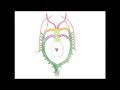 Medical Embryology - Development of the Aortic Arches and Large Arteries
 Easy Ways to Remember Heart Embryology derivatives
 Introduction to Embryology - Fertilisation to Gastrulation (Easy to Understand)
 Early Development of the heart: Malformations & Overview – Embryology | Lecturio
 Embryology | Development of Fetal Circulation
 Embryology of the Female & Male Reproductive System II (Easy to Understand)
 Cardiogenesis animation
 Heart Tubes - Embryology
 Embryology of the CNS (Easy to Understand)