Embryology of the Lungs (Easy to Understand)
The development of the lungs explained in less than 10 minutes.
If you are completely new to embryology and you want to understand it quickly, this should be the first video you watch:
- https://youtu.be/l5gUARhXWTY
Post any questions you have about the video below, I read all the comments:
***PLEASE SUPPORT ME***
GoFundMe
https://www.gofundme.com/f/minass
Facebook
https://www.facebook.com/M1NA55/
Instagram
@m1.nass
@mi.nass
Email me:
m.inass@outlook.com
Summary for your notes:
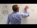
- Formation of the lung buds: at 4 weeks of gestation, the respiratory diverticulum (lung bud) appears as an outgrowth from the ventral foregut. Retinoic acid from surrounding mesoderm is the initiating factor (as it causes up-regulation of transcription factor TBX4) - not spoken about in video to keep things simple.
- Epithelium of the lung is derived from foregut.
- Connective tissue, muscle, and cartilage is from splanchnic mesoderm.
- Parietal pleura is from somatic mesoderm; and visceral pleura is from splanchnic mesoderm.
- Tracheoesophageal ridges separate the lung bud from the foregut, and fuse to form a tracheoesophageal septum.
- Lung buds form bronchial buds which form into the right and left main bronchi.
- Growth is caudal and lateral to fill the pericardioperitoneal canals.
10 tertiary bronchi (segmental) are developed on the right, and 8 in the left. These correspond to the bronchopulmonary segments in an adult lung.
- Terminal bronchioles divide to form respiratory bronchioles and these divide into alveolar ducts.
- The terminal sacs are initially cuboidal epithelium but the distal portion become squamous as the vascular supply grows near it - these are the Type 1 alveoli.
- Type 2 produce surfactant.
Congenital abnormalities:
- Esophageal atresia: abnormalities in partitioning of the oesophagus and trachea. Fistulas may also persist
- The three types spoken about in the video make up the most common types.
- The proximal blind ended esophagus, and a tracheoesophageal fistula to the distal part of the esopahgus is the most common type (90%).
- There are two other types which are much rarer (1% each) so it wasn't spoken about in the video. They include: (1) a proximal fistula, and a distal blind-ended esophagus, and (2) a fistula to the trachea from the esophagus both distally and proximally.
Видео Embryology of the Lungs (Easy to Understand) канала Dr. Minass
If you are completely new to embryology and you want to understand it quickly, this should be the first video you watch:
- https://youtu.be/l5gUARhXWTY
Post any questions you have about the video below, I read all the comments:
***PLEASE SUPPORT ME***
GoFundMe
https://www.gofundme.com/f/minass
https://www.facebook.com/M1NA55/
@m1.nass
@mi.nass
Email me:
m.inass@outlook.com
Summary for your notes:
- Formation of the lung buds: at 4 weeks of gestation, the respiratory diverticulum (lung bud) appears as an outgrowth from the ventral foregut. Retinoic acid from surrounding mesoderm is the initiating factor (as it causes up-regulation of transcription factor TBX4) - not spoken about in video to keep things simple.
- Epithelium of the lung is derived from foregut.
- Connective tissue, muscle, and cartilage is from splanchnic mesoderm.
- Parietal pleura is from somatic mesoderm; and visceral pleura is from splanchnic mesoderm.
- Tracheoesophageal ridges separate the lung bud from the foregut, and fuse to form a tracheoesophageal septum.
- Lung buds form bronchial buds which form into the right and left main bronchi.
- Growth is caudal and lateral to fill the pericardioperitoneal canals.
10 tertiary bronchi (segmental) are developed on the right, and 8 in the left. These correspond to the bronchopulmonary segments in an adult lung.
- Terminal bronchioles divide to form respiratory bronchioles and these divide into alveolar ducts.
- The terminal sacs are initially cuboidal epithelium but the distal portion become squamous as the vascular supply grows near it - these are the Type 1 alveoli.
- Type 2 produce surfactant.
Congenital abnormalities:
- Esophageal atresia: abnormalities in partitioning of the oesophagus and trachea. Fistulas may also persist
- The three types spoken about in the video make up the most common types.
- The proximal blind ended esophagus, and a tracheoesophageal fistula to the distal part of the esopahgus is the most common type (90%).
- There are two other types which are much rarer (1% each) so it wasn't spoken about in the video. They include: (1) a proximal fistula, and a distal blind-ended esophagus, and (2) a fistula to the trachea from the esophagus both distally and proximally.
Видео Embryology of the Lungs (Easy to Understand) канала Dr. Minass
Показать
Комментарии отсутствуют
Информация о видео
Другие видео канала
 Development of Respiratory System | Embryology
 Embryology of the Heart (Easy to Understand)
 THE DEVELOPMENT OF THE LUNGS AND THE RESPIRATORY SYSTEM-HUMAN EMBRYOLOGY-DR ROSE JOSE MD
 Embryology of the GIT II - Midgut (Easy to Understand)
 Oxygen - Haemoglobin Dissociation Curve - Physiology
 Respiratory | External Respiration: Ventilation Perfusion Coupling | Part 1
 Embryology of the Liver (Easy to Understand)
 Medical embryology - Difficult concepts of early development.mp4
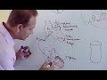 Ventricles and Aorticpulmonary Septum - Embryology
 Embryology of the Pancreas (Easy to Understand)
 Respiratory | Mechanics of Breathing: Pressure Changes | Part 1
 DEVELOPMENT OF THE GONADS-TESTIS AND OVARY-HUMAN EMBRYOLOGY -DR ROSE JOSE MD
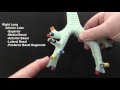 Naming the Segmental Bronchi
 Medical Embryology - Development of Body cavities, Intraembryonic coelom, and diaphragm
 Embryology of the Pharyngeal Arches (Easy to Understand)
 Cardiovascular | Electrophysiology | Intrinsic Cardiac Conduction System
 Respiratory System - Overview
 Embryology of the Eye (Easy to Understand)
 Embryology of the GIT III - Hindgut (Easy to Understand)
 Embryology of the Kidney (Easy to Understand)