Ultrasound Video showing Fetal anomalies, Clubfoot, Encephalocele, Kyphosis, and Placental Mass.
This video shows Fetal anomalies, including Clubfoot, Encephalocele, Kyphosis, and Placental Mass.
What can an ultrasound tell about a baby? Ultrasound can detect some types of physical birth defects. Examples of physical birth defects that may be found at 19 - 20 weeks are most cases of spina bifida, some serious heart defects, some kidney problems, absence of part of a limb, and some cases of cleft palate.
An encephalocele may be seen as a purely cystic mass or may contain echoes from herniated brain tissue. If the mass appears cystic, the meningocele component predominates, while a solid mass indicates predominantly an encephalocele. Larger encephaloceles may show accompanying microcephaly.
Fetuses with an encephalocele are likely to die before birth. Approximately 21 percent, or 1 in 5, are born alive. Of those born alive, only 50 percent will survive.
After 12 weeks' gestation: the spine is distorted in the sagittal or coronal view resulting in scoliosis. The hemivertebra appears as a triangular bony structure, smaller than a regular vertebra, acting as a wedge against the adjacent normal vertebras.
Scanning the fetus spine in the Sagittal plane, an anterior bulge is seen.
Kyphosis is a spinal disorder in which an excessive outward curve of the spine results in an abnormal rounding of the upper back. The condition is sometimes known as "roundback" or—in the case of a severe curve—as "hunchback." Kyphosis can occur at any age but is common during adolescence.
Kyphosis is most common in the thoracic spine, though it can also affect the cervical and lumbar spine. There are several causes of kyphosis in adults. The first is congenital, which means it is a condition present from birth. A congenital spine problem affects the development of the spine.
Congenital kyphosis is caused when something disrupts the normal development of the spine before birth. In many cases, two or more of the vertebrae fuse together. It's often unclear why certain children are affected in this way.
Clubfoot describes a range of foot abnormalities usually present at birth (congenital) in which your baby's foot is twisted out of shape or position. In clubfoot, the tissues connecting the muscles to the bone (tendons) are shorter than usual.
Congenital talipes equinovarus (CTEV), often also known as ‘club-foot’, is a common developmental disorder of the lower limb. It is defined as fixation of the foot in adduction, in supination and in varus, i.e. inclined inwards, axially rotated outwards, and pointing downwards. The calcaneus, navicular and cuboid bones are medially rotated in relation to the talus and are held in adduction and inversion by ligaments and tendons. The foot is supinated, but the front of the foot is pronated in relation to the back of the foot, causing cavus. Moreover, the first metatarsal is more plantarflexed.
One or both feet are rotated inward and downward. The affected foot and leg may be smaller in size compared to the other. Approximately 50% of cases of clubfoot affect both feet. Mostly it is not associated with other problems. The foot remains deformed without treatment, and the affected person walks on the sides of the feet and it may cause pain and difficulty in walking.
The exact cause is usually could not be identified but genetic and environmental factors are believed to be involved. In identical twins, if one fetus is affected, there is a 33% chance that the other one may be affected.
Clubfoot is mainly idiopathic, which means that the cause is unknown. Genetic factors are believed to play a major role, and some specific gene changes have been associated with it, but this is not yet well understood. It appears to be passed down through families. It is not caused by the fetus's position in the uterus.
Clubfoot is a fairly common birth defect in which the foot is twisted in and down. In severe cases, a club foot sufferer may become significantly disabled. If you find it difficult to work because of your club foot, you may be eligible for disability benefits under the Social Security program.
In fact, the condition does not bother the baby until the time he or she begins to stand and walk. This causes problems for the parts of the feet that are not normally walked on. The normal growth of the leg is also affected. Babies born with clubfoot should receive expert help shortly after birth.
Chorioangioma is a benign vascular malformation of the placenta and represents the most common primary tumor of the placenta.
Placental hemangioma. Chorioangioma is a benign tumor of the placenta. It is seen in approximately 0.5 to 1% pregnancies. It is mostly diagnosed ultrasonically in the second trimester of pregnancy. Large Chorioangioma is known to cause complications in pregnancy, while the smaller ones are asymptomatic.
Видео Ultrasound Video showing Fetal anomalies, Clubfoot, Encephalocele, Kyphosis, and Placental Mass. канала Saeed Ahmad
Показать
Комментарии отсутствуют
Информация о видео
Другие видео канала
 Ultrasound Video showing Cystic hygroma with Microcephaly.
 2nd Trimester OB Ultrasound Evaluation with Dr. Filly
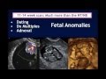 AIUM Webinar: Systematic Evaluation of the 11-14 Week Fetus, Touching on ISUOG Guidelines
 Hydrops fetalis, with Fetal Soft tissue edema, Ascites, and Pleural effusion.
 Ultrasound Scans & Prenatal Screening
 Fetale Echokardiographie
 The Basic Fetal Heart Scan
 Ultrasound Video showing multiple fetal anomalies.
 Ultrasound Video showing Hydatid Hepatic Cyst, Cholelithiasis and Hepatic masses.
 Fetal Measurements, Tips and Tricks
 Multiple findings including Gallbladder Stone, Large hepatic Mass, Vesical Growth, and BPH.
 Step by Step to Get Perfect 3D/4D Baby Image
 Ectopically placed Single Kidney.
 Nuchal translucency (NT)
 Ultrasound Video showing a case of club foot, also called talipes equinovarus (TEV).
 Fetal measurements and Doppler | How to do it !
 ANOMALY SCAN -Top 5 Tips | By Dr. Mukesh Gupta
 Bilateral Multiple Ovarian Cysts and Ovarian Mass.
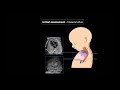 Fetal Echocardiography: Protocol and Technique
 Webinar Replay: How to diagnose Endometriosis on Ultrasound